
Page 57 - IJB-7-4
P. 57
Lin, et al.
A
B D
C E
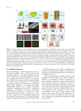
Figure 5. Schematic diagrams of other scaffold structures. (A) Schematic diagram of GML+TGL material mimic lotus pod scaffold
structure [106] . (From ref. [106] licensed under Creative Common Attribution-NonCommercial-NoDerivatives 4.0 International (CC BY-NC-
ND 4.0) (B) Schematic diagram of cell-carrying spring-like scaffold structure [107] . (Reprinted with permission from Gao Q, Liu Z, Lin
Z, et al., 3D Bioprinting of Vessel-like Structures with Multi-level Fluidic Channels, ACS Biomaterials Science and Engineering. 2017;
3(3):399-408. Copyright© 2017 American Chemical Society) (C) Schematic diagram of hexagonal mimic scaffold structure [108] . Reprinted
with permission from Ma X, Xin Q, Wei Z, et al. Deterministically patterned biomimetic human iPSC-derived hepatic model via rapid 3D
bioprinting. Proceedings of the National Academy of Sciences. 2016; 113(8):2206. (D) Light microscope images of multi-shape GelMA [109] .
Adapted from Xie, M., Yu, K., Sun, Y., Shao, L., Nie, J., Gao, Q., Qiu, J., Fu, J., Chen, Z., He, Y. Protocols of 3D Bioprinting of Gelatin
Methacryloyl Hydrogel Based Bioinks. J. Vis. Exp. (154), e60545, doi:10.3791/60545 (2019) (E) Schematic diagram of multi-layered helical
cylindrical scaffold structure [110] . Reprinted with permission from Xue J M, Feng C, Xia LG, et al. Assembly Preparation of Multilayered
©
Biomaterials with High Mechanical Strength and Bone-Forming Bioactivity. Chemistry of Materials. 2018; 30(14):4646-4657, Copyright
2018 American Chemical Society.
4. Composite function a cell-filled hydrogel as the bioink, cell printing can
With the development of tissue engineering technology, print any cells needed directly within the scaffold area to
prepare 3D cell scaffold structures with cell proliferation
biomedical scaffolds used in clinical practice are [122-127]
constantly updated [111-116]. 3D printed scaffolds are widely and differentiation .
used in regenerating tissues and organs such as skin, However, bioprinting of cellular structures faces
nerve, bladder, bone, and blood vessels [117-119] . However, significant obstacles, including the impact of different
it is still difficult to prepare ideal 3D printable scaffolds materials on maintaining mechanical properties at
that promote tissue regeneration [120] . 3D bioprinting, micro- and macro-scopic scales, achieving tissue designs
a recently developed biomanufacturing technology, with biological specificity, developing methods to
addresses this challenge by providing unprecedented obtain and expand functional cells from stem cells, and
manufacturing precision by a highly controllable connecting bioprinted tissues to the physiological vascular
mechanical manufacturing mechanism [121] . Bioprinting system. The initial success of clinical applications of
technology is controlled by a computer-aided design 3D bioprinting for the preparation of active tissues
system and can manufacture a variety of complex was attributed to the relatively simple geometry of the
microstructures layer by layer. Cell printing is one of prepared active tissues. Based on this perspective, we
the more widely used 3D bioprinting methods that provide an overview of recent advances in bio-3D printed
can overcome the drawbacks of conventional cell-free active scaffolds and a generalized enumeration of their
printed 3D scaffolds by loading cells in bioink. Using main functions.
International Journal of Bioprinting (2021)–Volume 7, Issue 4 53