
Page 54 - IJB-7-4
P. 54
Filament Structure, 3D printing, Bone Repair Scaffolds
achieve higher osteoinductivity of bioceramic materials, parallel. The advantage of the hollow structure is that the
Alksne et al. prepared two bilayer scaffolds, that is, PLA scaffold has large porosity to facilitate the growth and
[93]
+ HA and PLA + BG (Figure 2D); PLA + BG scaffolds flow of osteoblasts and growth factors, transport nutrients
were 15% more absorbent than other controls, provided and load drugs, and its internal structure also provides a
better nutrient and protein uptake, and induced the earliest suitable space for the development of vascular growth.
onset of ALP activity and the highest cellular activity, and Feng et al. successfully prepared a lotus root-like
[95]
a large amount of protein deposition was found on the bone repair scaffold with parallel multichannel structure
surface of PLA + BG scaffold. Due to the high process (channel-struts-packed, 1-4CSP) using Mg yellow
ability of cell-carrying bioceramic scaffolds, Kim et al. feldspar (Figure 3A). The porosity (80%) and specific
[94]
prepared α-TCP/COL scaffolds with ceramic volume surface area (~3.86 m g ) of the mimetic material were
2 −1
fraction over 70% by modulating printing parameters significantly higher, and micro-computed tomography
using preosteoblasts (Figure 2E), which had a higher results showed that the BV/TV values were significantly
elastic modulus (~0.55 MPa) compared to the control higher in the 3CSP group (12.6%) after 12 weeks of
group and a cell survival rate of over 91% (within 4 h), implantation. They found that the porous scaffold is more
concluding that cell-laden ceramic scaffold is a potentially suitable for cell delivery and regeneration of large bone
viable solution for bone regeneration. defects. The complexity of the hierarchical structure,
the mechanical properties required and the diversity of
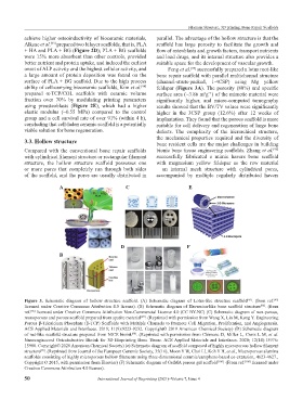
3.3. Hollow structure bone resident cells are the major challenges in building
[96]
Compared with the conventional bone repair scaffolds bionic bone tissue engineering scaffolds. Zhang et al.
with cylindrical filament structure or rectangular filament successfully fabricated a mimic havers bone scaffold
structure, the hollow structure scaffold possesses one with magnesium yellow feldspar as the raw material
or more pores that completely run through both sides – an internal mesh structure with cylindrical pores,
of the scaffold, and the pores are usually distributed in accompanied by multiple regularly distributed havers
A C E
D F
B
Figure 3. Schematic diagram of hollow structure scaffold. (A) Schematic diagram of Lotus-like structure scaffold . (from ref.
[95]
[95]
licensed under Creative Commons Attribution 4.0 license). (B) Schematic diagram of Haversian-like bone scaffold structure . (from
[96]
ref. licensed under Creative Commons Attribution Non-Commercial License 4.0 (CC BY-NC) (C) Schematic diagram of non-porous,
[96]
monoporous and porous scaffold prepared from apatite material . (Reprinted with permission from Wang X, Lin M, Kang Y. Engineering
[97]
Porous β-Tricalcium Phosphate (β-TCP) Scaffolds with Multiple Channels to Promote Cell Migration, Proliferation, and Angiogenesis.
ACS Applied Materials and Interfaces. 2019; 11(9):9223-9232. Copyright© 2019 American Chemical Society) (D) Schematic diagram
of nut-like scaffold structure prepared from NICE bioink . (Reprinted with permission from Chimene D, Miller L, Cross L M, et al.
[98]
Nanoengineered Osteoinductive Bioink for 3D Bioprinting Bone Tissue. ACS Applied Materials and Interfaces. 2020; 12(14):15976-
15988. Copyright© 2020 American Chemical Society) (e) Schematic diagram of scaffold composed of highly microporous hollow filament
structure . (Reprinted from Journal of the European Ceramic Society, 35(16), Moon Y W, Choi I J, Koh Y H, et al., Macroporous alumina
[99]
scaffolds consisting of highly microporous hollow filaments using three-dimensional ceramic/camphene-based co-extrusion, 4623-4627.,
Copyright © 2015, with permission from Elsevier) (F) Schematic diagram of GelMA porous gel scaffold [100] . (From ref. [100] licensed under
Creative Commons Attribution 4.0 license).
50 International Journal of Bioprinting (2021)–Volume 7, Issue 4