
Page 52 - IJB-7-4
P. 52
Filament Structure, 3D printing, Bone Repair Scaffolds
Table 2. Summary of the characteristic of different structures in 3D bioprinted biodegradable bone repair scaffolds
Structure Features References
Classic Structure Easy parameter adjustment, simple preparation process, [82-86]
secondary processing potential
Double layer structure Effectively improve the mechanical strength of the stent [5],[88-91]
and enrich the function of the stent
Hollow structure Large pore structure for nutrient delivery and drug [92-97]
loading, providing space for blood vessel growth
Core-shell structure Ensures material independence, adjustable scaffold [98-101]
degradation rate
Bionic structures and Free shape customization based on defects, easy to load [103-107]
others cells
A B C
D E F
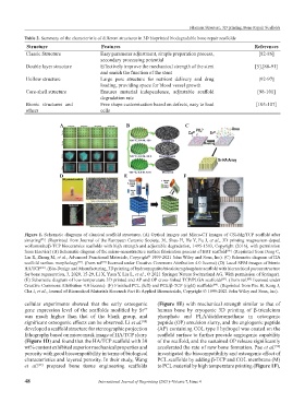
Figure 1. Schematic diagrams of classical scaffold structures. (A) Optical images and Micro-CT images of CSi-Mg/TCP scaffold after
sintering . (Reprinted from Journal of the European Ceramic Society, 36, Shao H, He Y, Fu J, et al., 3D printing magnesium-doped
[85]
wollastonite/β-TCP bioceramics scaffolds with high strength and adjustable degradation, 1495-1503, Copyright (2016), with permission
from Elsevier) (B) Schematic diagram of the micro-nanostructure surface fabrication process of BRT scaffold . (Reprinted from Deng C,
[86]
Lin R, Zhang M, et al., Advanced Functional Materials, Copyright 1999-2021 John Wiley and Sons, Inc). (C) Schematic diagram of HA
©
scaffold surface morphology . (from ref licensed under Creative Commons Attribution 4.0 license) (D) Local SEM images of bionic
[87]
[87]
HA/TCP . (Bio-Design and Manufacturing, 3D printing of hydroxyapatite/tricalcium phosphate scaffold with hierarchical porous structure
[88]
for bone regeneration, 3, 2020, 15-29, Li X, Yuan Y, Liu L, et al., © 2021 Springer Nature Switzerland AG. With permission of Springer).
(E) Schematic diagram of low-temperature 3D printed and AP and OP cross-linked TCP/PLGA scaffolds . (from ref. licensed under
[89]
[89]
Creative Commons Attribution 4.0 license). (F) Finished PCL (left) and PCL/β-TCP (right) scaffolds . (Reprinted from Pae H, Kang J,
[90]
Cha J, et al., Journal of Biomedical Materials Research Part B: Applied Biomaterials, Copyright © 1999-2021 John Wiley and Sons, Inc).
cellular experiments showed that the early osteogenic (Figure 1E) with mechanical strength similar to that of
gene expression level of the scaffolds modified by Sr human bone by cryogenic 3D printing of β-tricalcium
2+
was much higher than that of the blank group, and phosphate and PLA/dichloromethane in osteogenic
significant osteogenic effects can be observed. Li et al. peptide (OP) emulsion slurry, and the angiogenic peptide
[88]
developed a scaffold structure for stereographic projection (AP) containing COL type I hydrogel was coated on the
lithography based on micro mask image of HA/TCP slurry scaffold surface to further provide angiogenic capability
(Figure 1D) and found that the HA/TCP scaffold with 30 of the scaffold, and the sustained OP release significantly
wt% content exhibited superior mechanical properties and accelerated the rate of new bone formation. Pae et al.
[90]
porosity with good biocompatibility in terms of biological investigated the biocompatibility and osteogenic effect of
characteristics and layered porosity. In their study, Wang PCL scaffolds by adding β-TCP and COL membrane (M)
et al. prepared bone tissue engineering scaffolds to PCL material by high temperature printing (Figure 1F),
[89]
48 International Journal of Bioprinting (2021)–Volume 7, Issue 4