
Page 92 - IJB-7-4
P. 92
3D Printing Osteochondral Scaffold
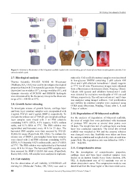
Figure 1. Schematic illustration of the bilayered scaffold loaded with transforming growth factor-β and bone morphogenetic protein-2 for
osteochondral repair.
2.7. Rheological analysis stain cells. Cell-scaffold construct samples were incubated
in low-glucose DMEM containing 2 μM calcein AM
Thermo Scientific HAAKE MARS 40 Rheometer (live) and 4 μM ethidium homodimer-1 (dead) reagents
(Waltham, MA, USA) was used to investigate rheological at 37°C for 45 min. Fluorescence images were obtained
properties fitted with 25 mm parallel geometry. Frequency- from a fluorescence microscope (Zeiss, Nanjing, China).
dependent loss modulus (G’’), storage modulus (G’), and Calcein AM (green) and ethidium homodimer-1 (red)
dynamic viscosity of DCM/SF and DBM/SF hydrogels were detected by excitation wavelengths of 495 nm and
were determined by the frequency sweep in the shear rate 560 nm, respectively. The cell survival rate at 1 and 3 days
range of 0.1~100 Hz at 15℃. was analyzed using ImageJ software. Cell proliferation
2.8. Growth factor releasing and viability in construct samples were examined using
CCK8 assay (Beyotime, Nanjing, China) after 1, 4, and
To investigate release of growth factors, cartilage layer 7 days of culture.
and bone layer construct samples were encapsulated with
2 μg/mL TGF-β1 and 2 μg/mL BMP-2, respectively. To 2.10. Degradation of 3D bilayered scaffolds
evaluate the release rate of TGF-β1, pre-weighed cartilage For the analysis of degradation of bilayered scaffolds,
layer samples were rinsed with 2 ml PBS solutions the rates of weight loss were performed with treatment
containing 0.05% EDTA, 0.1% heparin, 0.02% sodium of protease XIV enzyme at several time points over
azide, and 0.1% BSA at 37°C. The PBS solution was 24 days. The weight loss test of cartilage layer and bone
replenished and harvested every 48 h for 21 days. The layer was conducted separately. The initial dry printed
harvested PBS samples were then assessed by TGF-β1 scaffold was weighed as W0 and the enzyme solution
ELISA Kit assay (PeproTech, RH, USA). To evaluate the was changed every day. Scaffolds were taken out from
release rate of BMP-2, pre-weighed bone layer samples the enzyme solution and weighed at dry state at each time
were rinsed with 2 ml PBS solutions containing 0.05% point (Wd). The degradation rate was defined as 100% ×
EDTA, 0.1% heparin, 0.02% sodium azide, and 0.1% BSA (W0 – Wd)/W0.
at 37°C. The PBS solution was replenished and harvested
every 48 h for 14 days. The harvested PBS samples were 2.11. Comprehensive stress
then assessed by BMP-2 ELISA Kit assay (PeproTech, For the investigation of comprehensive properties,
Rocky Hill, USA). All samples were analyzed in triplicate. cartilage layer and bone layer construct samples were
2.9. Cell viability loaded on an Instron Tensile Force Tester (Instron, HW,
UK). A displacement rate of 0.1 mm/min was set to
For the observation of cell viability, LIVE/DEAD cell obtain stress-strain curve. The compression modulus was
staining kit (Molecular Probes, OR, USA) was used to determined from the linear region of the stress-strain curve.
88 International Journal of Bioprinting (2021)–Volume 7, Issue 4