
Page 94 - IJB-7-4
P. 94
3D Printing Osteochondral Scaffold
A B
C D
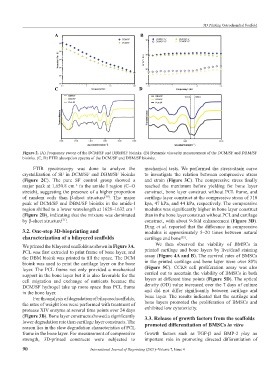
Figure 2. (A) Frequency sweep of the DCM/SF and DBM/SF bioinks. (B) Dynamic viscosity measurement of the DCM/SF and DBM/SF
bioinks. (C, D) FTIR absorption spectra of the DCM/SF and DBM/SF bioinks.
FTIR spectroscopy was done to analyze the mechanical tests. We performed the stress-strain curve
crystallization of SF in DCM/SF and DBM/SF bioinks to investigate the relation between compressive stress
(Figure 2C). The pure SF control group showed a and strain (Figure 3C). The compressive stress finally
major peak at 1,650.8 cm in the amide I region (C=O reached the maximum before yielding for bone layer
−1
stretch), suggesting the presence of a higher proportion construct, bone layer construct without PCL frame, and
of random coils than β-sheet structure . The major cartilage layer construct at the compressive stress of 310
[50]
peak of DCM/SF and DBM/SF bioinks in the amide-I kpa, 47 kPa, and 44 kPa, respectively. The compressive
region shifted to a lower wavelength at 1628–1632 cm modulus was significantly higher in bone layer construct
−1
(Figure 2D), indicating that the mixture was dominated than in the bone layer construct without PCL and cartilage
by β-sheet structure . construct, with about 9-fold enhancement (Figure 3D).
[51]
Ding et al. reported that the difference in compressive
3.2. One-step 3D-bioprinting and modulus is approximately 5–20 times between natural
characterization of a bilayered scaffolds cartilage and bone .
[52]
We printed the bilayered scaffolds as shown in Figure 3A. We then observed the viability of BMSCs in
PCL was first extruded to print frame of bone layer, and printed cartilage and bone layers by live/dead staining
the DBM bioink was printed to fill the space. The DCM assay (Figure 4A and B). The survival rates of BMSCs
bioink was used to print the cartilage layer on the bone in the printed cartilage and bone layer were over 80%
layer. The PCL frame not only provided a mechanical (Figure 5C). CCK8 cell proliferation assay was also
support in the bone layer but it is also favorable for the carried out to ascertain the viability of BMSCs in both
cell migration and exchange of nutrients because the layers at different time points (Figure 5D). The optical
DCM/SF hydrogel take up more space than PCL frame density (OD) value increased over the 7 days of culture
in the bone layer. and did not differ significantly between cartilage and
For the analysis of degradation of bilayered scaffolds, bone layer. The results indicated that the cartilage and
the rates of weight loss were performed with treatment of bone layers promoted the proliferation of BMSCs and
protease XIV enzyme at several time points over 24 days exhibited low cytotoxicity.
(Figure 3B). Bone layer constructs showed a significantly 3.3. Release of growth factors from the scaffolds
lower degradation rate than cartilage layer constructs. The promoted differentiation of BMSCs in vitro
reason lies in the slow degradation characteristics of PCL
frame in the bone layer. For measurement of compressive Growth factors such as TGF-β and BMP-2 play an
strength, 3D-printed constructs were subjected to important role in promoting directed differentiation of
90 International Journal of Bioprinting (2021)–Volume 7, Issue 4