
Page 95 - IJB-7-4
P. 95
Zhang, et al.
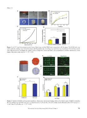
A B
C D
Figure 3. (A) PCL was first extruded to print frame of bone layer, and the DBM bioink was printed to fill the space. The DCM bioink was
used to print the cartilage layer on the bone layer. (B) Degradation rate of cartilage layer and bone layer in protease XIV. (C) Stress-strain
curve that was used to investigate the relation between compressive stress and strain. (D) Comprehensive modulus calculated by linear
region of the stress-strain curve (n = 3; *P < 0.05).
A
B
A
C D
Figure 4. Viability of BMSCs in the printed constructs. Fluorescence microscopy findings of the LIVE/DEAD assay of BMSCs cultured in
the (A) cartilage layer and (B) bone layer. (C) Quantification of cell viability. (D) Cell viability results obtained by the CCK-8 assay on 1,
4, and 7 days of cell culture (n = 3; *P < 0.05).
International Journal of Bioprinting (2021)–Volume 7, Issue 4 91