
Page 25 - IJB-8-1
P. 25
Tian, et al.
customizable, so that we can fabricate hands of various
shapes and sizes easily; second, our robotic hands
should have the similar functionality of the human hand,
especially in completing different grasping gestures. To
this end, we propose a multi-layer deformable design
of robotic hands, which mainly consists of (i) a layer of
silicone skin, (ii) a layer of 3D printed tissues, (iii) a layer
of 3D printed bones, and (iv) an underactuated system.
Specifically, given a real human hand as the target,
we first obtained its surface model through a 3D scanner.
The skin layer could be de-molded directly from the target
hand to mimic the appearance of the target hand. Then,
we proposed a fast template matching method to obtain
the corresponding 3D bone models based on the surface
model (Section 2.3). After that, an effective concentric
tube structure was adopted to construct the tissue layer
(Section 2.4). The tissue layer serves as the intermediate
layer between the skin and bones and is made of elastic
materials to allow the high deformability of our robotic
hands. At last, we adopted a low-cost cable-driven system
to provide our robotic hands with the mobility and
stability (Section 2.6).
Compared with conventional single layer/structure
robotic hands [3,13,15,25,27] , our multi-layer design is
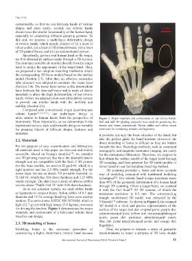
more similar to human hands from the perspective of Figure 1. Major materials and components of our robotic hands.
biomimetic. More importantly, as we demonstrate in the Soft and stiff 3D printing materials were used for producing the
experiment section, such a deformable design is versatile tissues and bones, respectively. Nylon cables and rubber bands
for grasping objects of different shapes, textures, and were used for simulating tendons and ligaments.
materials. it provides not only the basic structure of the hand, but
2.2. Materials also the perfect guide for hand motions. However, the
direct modeling of bones is difficult as they are hidden
For the purpose of easy customization and fabrication, beneath the skin. Radiology methods, such as computed
all materials used in this paper are low-cost and widely tomography and magnetic resonance imaging, are costly
accessible. Based on Young’s modulus , we consider for the customized fabrication. Therefore, we propose to
[18]
two 3D printing materials that have the desirable tensile first obtain the surface model of the target hand through
strength and are compatible with the form 2 3D printer. 3D scanning, and then generate the 3D mesh models of
For the bone models, we used rs-f2-gpcl-04, which is a bones based on our fast template matching method.
rigid material and has 2.8 GPa tensile strength. For the 3D scanning provides a faster and more accurate
tissue layer, we use an elastic 3D printable material, rs- way of modeling compared with traditional modeling
f2-elcl-01, which has 50A shore hardness and 3.23 MPa techniques . Our robotic hand design maintains more
[28]
tensile strength. The skin layer is made of silicone rubber than 90% of the geometric information of a human hand
and we chose “PlatSil Gel 10” with 10A shore hardness. through 3D scanning. Given a target hand, we scanned
As to our actuation system, we used rubber bands it with the Go! Scan 50 3D scanner, of which the
TM
as ligaments to connect bones. Nylon cables of 0.3 mm maximum resolution is 0.5 mm. The corresponding
diameter and 10 lbs average breaking force were used as triangle mesh-based 3D model was extracted by the
tendons. The servo motor, HITEC HS-5070HM, which is VXmodel software. As shown in Figure 2, the scanned
TM
light (12.7 g) yet with high torque (3.6 kg/cm), was used 3D model is a vivid and precise representation of the
for driving the tendons. Figure 1 demonstrates the major surface of the target (red dot: original point; purple dot:
materials and components of a fabricated robotic hand carpometacarpal joint; yellow dot: metacarpophalangeal
based on our design. joints; green dot: proximal interphalangeal joints;
blue dot: distal interphalangeal joints; and orange dot:
2.3. 3D modeling of bones fingertips).
Modeling bones is the necessary procedure of Next, we propose to estimate a series of geometric
customizing a highly biomimetic robotic hand because transformations, to match a template of 3D bone models
International Journal of Bioprinting (2022)–Volume 8, Issue 1 11