
Page 141 - IJB-8-3
P. 141
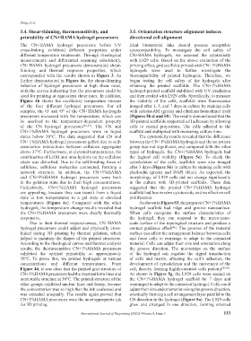
Dong, et al.
           3.4. Shear-thinning, thermosensitivity, and         3.5. Orientation structure alignment induces
           printability of CN+HAMA hydrogel precursors         directional cell alignment
           The  CN+HAMA  hydrogel  precursors  before  UV      Ideal  biomaterial  inks should possess acceptable
           cross-linking  exhibited  different  properties  under   cytocompatibility.  To investigate  the cell safety of
           different  temperature  treatments. Through  rheological   CN+HAMA  hydrogels,  we  assessed  the  cytotoxicity
           measurements  and  differential  scanning  calorimetry,   with L929 cells. Based on the above evaluation of the
           CN+HAMA  hydrogel  precursors  demonstrated  shear-  printing effect, grid scaffolds printed with CN+1%HAMA
           thinning and thermal response properties, which     hydrogels were used to further investigate  the
           corresponded with the results shown in Figure 3. As   biocompatibility  of printed hydrogels.  Therefore,  we
           further demonstrated in Figure 4a, the shear-thinning   began testing the cell safety of the hydrogels after
           behavior of hydrogel precursors at high shear rates,   obtaining  the  printed  scaffolds.  The  CN+1%HAMA
           with the curves indicating that the precursors could be   hydrogel-printed scaffold stabilized with UV irradiation
           used for printing at equivalent shear rates. In addition,   and then seeded with L929 cells. Specifically, to measure
           Figure  4b shows the oscillatory temperature sweeps   the  viability  of  the  cells,  scaffolds  were  fluorescence
           of  the  four  different  hydrogel  precursors.  For  all   imaged after 1, 3, and 7 days in culture by staining cells
           samples, the G’ and G″ of the CN+HAMA hydrogels     with calcein-AM (green) and ethidium homodimer (red)
           precursors increased with the temperature, which can   (Figures 5b-d and S5). The results demonstrated that the
           be ascribed to the temperature-dependent property   3D-printed scaffolds supported cell adhesion by allowing
           of the CN hydrogel precursors [42,43] .  The CN and   cells  to extend projections.  The  cells  adhered  to  the
           CN+1%HAMA  hydrogel  precursors  were  in  liquid   scaffold and multiplied with increasing culture time.
           states below 30°C.  The data suggested that CN and      The cytotoxicity results revealed that the difference
           CN+1%HAMA hydrogel precursors gelled due to self-   between the CN+1%HAMA hydrogels and the no pattern
           association interactions between cellulose aggregates   group was not significant, and compared with the other
           above 37°C. Furthermore, at elevated temperatures, the   types  of  hydrogels,  the  CN+1%HAMA  hydrogels  had
           combination of LiOH and urea hydrate on the cellulose   the  highest  cell  viability  (Figure  5e).  To  check  the
           chain was disturbed. Due to the self-binding force of   cytoskeleton  of  the  cells,  scaffolds  were  also  imaged
           cellulose, cellulose molecules connected, forming a   after 3 days (Figure S6) in culture by staining cells with
           network  structure.  In  addition,  the  CN+3%HAMA   phalloidin  (green)  and  DAPI  (blue).  As  expected,  the
           and  CN+5%HAMA  hydrogel  precursors  were  both    morphology  of  L929  cells  did  not  change  significantly
           in the gelation state due to their high concentrations.   in  the  culture  with  3D-printed  scaffolds.  These  data
           Collectively,  CN+1%HAMA  hydrogel  precursors      suggested  that  the  printed  CN+1%HAMA  hydrogel
           are appealing, because they can transit from a liquid   scaffold had low in vitro cytotoxicity and no effect on cell
           state at low temperatures to a gel state at elevated   proliferation.
           temperatures  (Figure  4c).  Compared  with  the  other   As shown in Figure S3, the prepared CN+1%HAMA
           hydrogels, the temperature change results revealed that   hydrogel  scaffold  had  ridge  and  groove  nanosurface.
           the  CN+1%HAMA  precursors  were  clearly  thermally   When  cells  recognize  the  surface  characteristics  of
           responsive.                                         the  hydrogel,  they  can  respond to the  micro-nano-
               Due  to  their  thermal  responsiveness,  CN+HAMA   scale surface of the topological structure and produce a
           hydrogel  precursors could  adjust and  physically  cross-  contact  guidance  effect . The grooves of the material
                                                                                   [44]
           linked  during 3D printing  by thermal  gelation,  which   surface can affect the arrangement balance between cells
           helped to maintain the shapes of the printed structures.   and  force  cells  to  rearrange  to  adapt  to  the  contacted
           According to the rheological curves and thermal analysis   material. Cells can adjust their size and orientation along
           results,  the  thermosensitive  CN+1%HAMA  precursors   the groove direction.  The microstrips on the surface
           exhibited  the optimal  printability  at approximately   of the hydrogel can regulate the signal transduction
           30°C.  To prove  this,  we printed  hydrogels  at  various   of  cells  and  matrix,  affecting  the  cell’s  adhesion,  the
           concentrations  and  different  temperatures.  From   development  of cytoskeleton  and the  movement  of the
           Figure 4d, it was clear that the printed grid structure of   cell, thereby forming highly-oriented cells patterns [45,46] .
           CN+1%HAMA precursors had the most uniform lines and   As shown in Figure 5g, the L929 cells were seeded on
           most stable structure at 30°C. The printed structure of the   the  CN+1%HAMA  hydrogel  scaffold  for  7  days  and
           other groups exhibited unclear lines and forms, because   rearranged to adapt to the contacted hydrogel. Cells could
           the concentration was so high that the ink coalesced and   adjust their size and orientation along the groove direction,
           was extruded  unequally.  The  results  again  proved  that   eventually forming a cell arrangement layer parallel to the
           CN+1%HAMA precursors were the most appropriate ink   CN direction in the hydrogel (Figure 5a). The L929 cells
           for 3D printing.                                    grew and arranged in one direction, forming oriented

                                       International Journal of Bioprinting (2022)–Volume 8, Issue 3       133