
Page 144 - IJB-8-3
P. 144
Dual-Response Composite Hydrogels
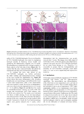
A
B
C
D
Figure 6. Biosafety and characterization of CN+1%HAMA hydrogels in Kunming Mouse models. (A) Schematic illustration of implanting
hydrogel samples in Kunming mouse models. Subcutaneously implanted hydrogels were explanted with the surrounding tissue after 1, 3, or
10 weeks. Sections were subjected to H&E staining (B) and stained red for CD3 (C) or CD68 (D) antigens.
safety of CN+1%HAMA hydrogels. The in vivo biosafety demonstrated that the immunoreaction was greatly
of CN+1%HAMA hydrogels was tested by implanting reduced after 3 weeks. The images show little signal of
hydrogel samples in Kunming mouse models and both kinds of cells, which indicated that the inflammatory
monitoring the inflammatory response over 10 weeks. response was gone and that CN+1%HAMA hydrogels
We subcutaneously implanted sterile hydrogel samples or do not cause tissue damage in vivo. Therefore, it is
blank controls in the back skin of mice (Figure 6a). We reasonable to assume that the biodegradability and
ensured that the mice remained healthy throughout the biosafety of CN+1%HAMA hydrogels could allow
experiment until they were sacrificed for euthanasia. them to be used in tissue engineering and regenerative
To evaluate the toxic side effects of the medicine.
CN+1%HAMA hydrogels, we, further, performed
the H&E staining and immunofluorescence staining 4. Conclusions
of the tissue around the implantation site. Figure 6b In this study, we successfully developed a novel CN+HAMA
clearly shows that many inflammatory cells, including hydrogels with thermo- and UV light-responsive
lymphocytes and macrophages, had infiltrated the tissue capabilities. The nanocellulose was thermosensitive,
1 week after implantation. Only a few inflammatory with a fluid state at low temperatures and a gel state at
cells were found at 3 weeks. No toxicity or inflammatory body temperature (>30°C). This reversible change allows
cells were observed in the implantation site at 10 weeks the hydrogels to gel in situ at temperatures >30°C while
(n = 5). In addition, Figure 6c and 6d show the results maintaining stability at low temperatures (even below
of immunofluorescence staining of T-cells with CD3 0°C). In addition, the incorporation of a UV-cross-linked
antigen and macrophages with CD68 antigen, both HAMA polymer network could improve the temperature-
of which were colored red. The results were consistent responsiveness of the system. Furthermore, the CNs could
with those of H&E staining. T-cells and macrophages be aligned directionally due to the shear stress-induced
were observed after 1 week. This is because the immune by the extrusion process. As a result, the cells seeded on
systems of the mice responded to foreign implants after the printed scaffold grew directionally, which indicated
1 week. Compared to 1 week, the T-cell and macrophage that the hydrogels could be used as potential candidate
signals were greatly reduced after 3 weeks, which ink for the fabrication of oriented soft-tissue mimics.
136 International Journal of Bioprinting (2022)–Volume 8, Issue 3