
Page 143 - IJB-8-3
P. 143
Dong, et al.
A
B F
C G
D H
E I
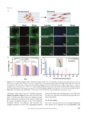
Figure 5. CN+1%HAMA hydrogels induce directional growth of L929 cells. (A) Schematic diagram of the growth process of cells on
the surface of oriented fibers. (B) Day 1, (C) Day 3, and (D) Day 7 fluorescence images of printed scaffolds seeded with L929 cells with
live/dead stain. The scale bar is 500 μm. (E) Cell viability comparison between various hydrogels on different days (Days 1, 3, and 7),
measured by a CCK-8 assay. (*P < 0.05, **P < 0.01). (F) The growth process of L929 cells seeded on culture dishes without treatment.
(G) The growth process of cells seeded on printed CN+1%HAMA hydrogel scaffolds. (H) A zoomed-in view of the frame column section in
Figure 6g. (I) Orientation angle distribution of L929 cells on CN+1%HAMA hydrogel scaffolds after culturing for 7 days.
cell patterns when cultured on CN+1%HAMA hydrogels functions for improving cell alignment in vitro. This work
(Figure 5g and h). Figure 5f shows that the L929 cells provides new structure-oriented hydrogel applications in
grew in all directions when they were cultured on petri tissue engineering.
dishes without any treatment. These results verified that
the unique orientation structure of the CN+1%HAMA 3.6. In vivo safety
hydrogels affected cell adhesion and promoted the An in vitro cell assay and an in vivo animal experiment
arranged orientation of L929 cells, suggesting potential were carried out to illustrate the biocompatibility and
International Journal of Bioprinting (2022)–Volume 8, Issue 3 135