
Page 153 - IJB-8-3
P. 153
Tang, et al.
A C
B
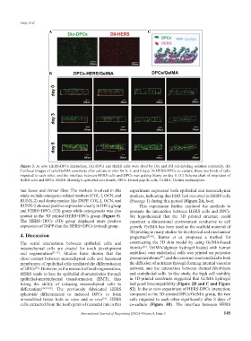
Figure 3. In vitro HERS-DPCs interaction. (A) DPCs and HERS cells were dyed by Dio and Dil cell-labeling solution separately. (B)
Confocal images of cells/GelMA constructs after culture in vitro for 0, 3, and 8 days. In HERS-DPCs co-culture, these two kinds of cells
migrated to each other, and the interface between HERS cells and DPCs was getting blurry on day 8. (C) Schema chart of migration of
HERS cells and DPCs. HERS: Hertwig’s epithelial root sheath, DPCs: Dental papilla cells, GelMA: Gelatin methacrylate.
had fewer and thinner fiber. The markers involved in this experiment expressed both epithelial and mesenchymal
study include osteogenic-related markers (COL-Ⅰ, OCN, and markers, indicating that EMT had occurred in HERS cells
RUNX-2) and dentin marker like DSPP. COL-Ⅰ, OCN, and (Passage 1) during this period (Figure 2A, b-c).
RUNX-2 showed positive expression clearly in DPCs group This experiment further explored the methods to
and HERS+DPCs (3D) group while osteogenesis was also promote the interaction between HERS cells and DPCs.
spotted in the 3D printed HERS+DPCs group (Figure 5). We hypothesized that the 3D printed structure could
The HERS+DPCs (3D) group displayed more positive construct a dimensional environment conducive to cell
expression of DSPP than the HERS+DPCs (mixed) group. growth. GelMA has been used as the scaffold material of
3D printing in many studies for its physical and mechanical
4. Discussion properties [32,41] . Barros et al. proposed a method for
The serial interactions between epithelial cells and constructing the 3D skin model by using GelMA-based
[25]
mesenchymal cells are crucial for tooth development bioinks . GelMA/alginate hydrogel loaded with human
and regeneration [35-37] . Studies have shown that the umbilical vein endothelial cells was printed on polyester
[41]
close contact between mesenchymal cells and basement porous membrane , and the construct was beneficial to both
membranes of epithelial cells mediated the differentiation the diffusion of nutrients through forming internal vascular
of DPCs . However, in the research of tooth regeneration, network and the interaction between dermal fibroblasts
[38]
HERS tends to lose its epithelial characteristics through and endothelial cells. In this study, the high cell viability
epithelial-mesenchymal transformation (EMT), thus in 3D-printed constructs suggested that GelMA hydrogel
losing the ability of inducing mesenchymal cells to had good biocompatibility (Figure 2B and C and Figure
differentiate [26,39,40] . The previously fabricated HERS S2). In the in vitro experiment of HERS-DPCs interaction,
spheroids differentiated or induced DPCs to form compared to the 3D-printed DPCs/GelMA group, the two
mineralized tissue both in vitro and in vivo . HERS cells migrated to each other significantly after 8 days of
[10]
cells extracted from the tooth germ of neonatal rats in this co-culture (Figure 3B). The interface between HERS
International Journal of Bioprinting (2022)–Volume 8, Issue 3 145