
Page 155 - IJB-8-3
P. 155
Tang, et al.
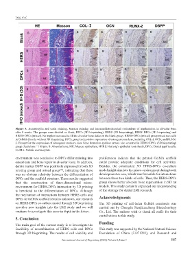
Figure 5. Hematoxylin and eosin staining, Masson staining and immunohistochemical evaluations of implantation in alveolar bone
after 8 weeks. The groups were divided as blank, DPCs (3D bioprinting), HERS (3D bioprinting), HERS+DPCs (3D bioprinting) and
HERS+DPCs (mixed). No implant was used to fill the alveolar bone defect in the blank group. HERS+DPCs (mixed) group mixed two cells
in GelMA directly without 3D bioprinting. DPCs group had positive expression of osteogenic markers, including COL-I, OCN, and RUNX-
2. Except for the expression of osteogenic markers, new bone formation (yellow arrow) also occurred in HERS+DPCs (3D bioprinting)
group. Scale bars = 100 μm. B: Alveolar bone, ME: Mucosa epithelium, HERS: Hertwig’s epithelial root sheath, DPCs: Dental papilla cells,
GelMA: Gelatin methacrylate.
environment was conducive to DPCs differentiating into proliferation indicate that the printed GelMA scaffold
osteoblasts and bone repair in alveolar fossa. In addition, could provide adequate conditions for cell activities.
dentin marker DSPP was positively expressed in both 3D Besides, the constructed 3D HERS-DPCs co-culture
printing group and mixed group , indicating that there model might simulate the micro-environment during tooth
[47]
was no obvious relativity between the differentiation of development in vivo, which was favorable for interactions
DPCs and the scaffold structure. These results suggested between these two kinds of cells. Thus, the HERS-DPCs
that the construction of three-dimensional micro- group shows better alveolar bone regeneration in SD rat
environment for HERS-DPCs interaction by 3D printing models. This study certainly expanded our understanding
is beneficial to the differentiation of DPCs. Although of the strategy for dental EMI research.
the mechanism of interactions between HERS cells and
DPCs in GelMA scaffold remains unknown, our research Acknowledgments
on HERS-DPCs co-culture model through 3D bioprinting The 3D printing of cell-laden GelMA constructs was
provides new insights into the EMI study and we will carried out by Chengdu Renjitiancheng Biotechnology
continue to investigate this issue in depth in the future. Co., Ltd. The authors wish to thank all staffs for their
contributions to this study.
5. Conclusion
The main goal of the current study is to investigate the Funding
feasibility of recombination of HERS cells and DPCs This study was supported by the National Natural Science
through 3D bioprinting. The results of cell viability and Foundation of China (31971281), and Research and
International Journal of Bioprinting (2022)–Volume 8, Issue 3 147