
Page 150 - IJB-8-3
P. 150
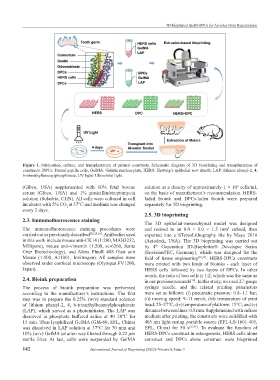
3D-bioprinted HERS-DPCs for Alveolar Bone Regeneration


           Figure  1.  Fabrication,  culture,  and transplantation  of printed  constructs.  Schematic  diagram  of 3D bioprinting  and  transplantation  of
           constructs. DPCs: Dental papilla cells, GelMA: Gelatin methacrylate, HERS: Hertwig’s epithelial root sheath, LAP: lithium phenyl-2, 4,
           6-trimethylbenzoylphosphinate, UV light: Ultraviolet light.

           (Gibco,  USA) supplemented  with 10% fetal  bovine   solution at a density of approximately 1 × 10  cells/mL
                                                                                                      6
           serum  (Gibco,  USA) and  1% penicillin/streptomycin   on the basis of manufacturer’s recommendation. HERS-
           solution (Solarbio, CHN). All cells were cultured in cell   laded  bioink  and DPCs-laden bioink  were  prepared
           incubator with 5% CO at 37°C and medium was changed   separately for 3D bioprinting.
                             2
           every 2 days.
                                                               2.5. 3D bioprinting
           2.3. Immunofluorescence staining
                                                               The 3D epithelial-mesenchymal model was designed
           The  immunofluorescence  staining  procedures  were   and resized to an 8.0 × 8.0 × 1.5 mm  cuboid, then
                                                                                                  3
           carried out as previously described [26,28,29] . Antibodies used   exported into a STereoLithography  file  by  Maya  2016
           in this work include mouse anti-CK14 (1:200, MAB3232,   (Autodesk,  USA).  The  3D bioprinting was carried out
           Millipore), mouse anti-vimentin (1:200, sc-6260, Santa   by 4   Generation 3D-Bioplotter® Developer Series
                                                                   th
           Cruz Biotechnology),  and Alexa FluoR 488 Goat anti   (EnvisionTEC, Germany), which was designed for the
           Mouse (1:500, A11001, Invitrogen). All  samples  were   field  of  tissue  engineering [30,31] . HERS-DPCs constructs
           observed under confocal microscope (Olympus FV1200,   were printed with two kinds of bioinks  -  each layer of
           Japan).                                             HERS cells followed by two layers of DPCs. In other
                                                               words, the ratio of two cells is 1:2, which was the same as
           2.4. Bioink preparation                             in our previous research . In this study, we used 27-gauge
                                                                                  [10]
           The process of bioink preparation was  performed    syringe needle, and the related printing parameters
           according  to  the  manufacturer’s  instructions.  The  first   were set as follows: (i) pneumatic pressure: 0.8–1.1 bar;
           step was to prepare the 0.25% (w/v) standard solution   (ii) moving speed: 9–11 mm/s; (iii) temperature of print
           of  lithium  phenyl-2,  4,  6-trimethylbenzoylphosphinate   head: 25–27℃; (iv) temperature of platform: 15°C; and (v)
           (LAP), which served as a photoinitiator. The LAP was   distance between lines: 0.8 mm. Supplemented with culture
           dissolved  in  phosphate  buffered  saline  at  40–50℃  for   medium after printing, the constructs were solidified with
           15 min. Then lyophilized GelMA (GM-60, EFL, China)   405 nm light-curing portable source (EFL-LS-1601-405,
           was dissolved in LAP solution at 37°C for 30 min and   EFL, China) for 30  s [25,32] .  To evaluate the function of
           10% (w/v) GelMA solution was filtered through 0.22 μm   HERS-DPCs construct in osteogenesis, HERS cells alone
           sterile  filter.  At  last,  cells  were  suspended  by  GelMA   construct and DPCs alone construct were bioprinted

           142                         International Journal of Bioprinting (2022)–Volume 8, Issue 3