
Page 24 - IJB-8-3
P. 24
Hydrogel based 3D-printing Bioinks for Cartilage Repair
acid (HA), alginate, collagen (COL), silk fibroin (SF),
and synthetic polymers such as gelatin methacryloyl
(GelMA) and polyethylene glycol (PEG) (Table 4) [11] .
This review focus on the properties of the above five
most commonly used hydrogels. We also discuss the
development and applications of such hydrogel-based
bioinks modified with functional additives. Finally,
challenges and future directions of hydrogels in the field
of cartilage regeneration are stated.
2. Overview of bioinks for 3D printed
cartilage engineering
The 3D-bioprinting technique applied in cartilage tissue
engineering usually contains three important elements,
i.e., cells, growth factors, and printed scaffolds, which
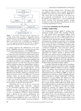
Figure 1. The general pathological process underlying OA.
Cartilage damage caused by injury or inflammation promotes are composed of various bioinks. Repair mechanisms of
the hypersecretion of pro-inflammatory cytokines such as IL-1β bioinks mainly involve two ways: (i) The printouts serve as
and IL-6, which then enhance the expression of MMPs including a temporary ECM environment to promote chondrogenesis
MMP-13 and ADAMTS such as ADAMTS-4 and ADAMTS-5 . and angiogenesis, leading to the generation of new cartilage
[6]
As a result, aggrecan proteoglycan and ECM COL are degraded, tissue; (ii) the engineered biomaterials replace the battered
leading to further deconstruction of damaged cartilage tissue. or lost cartilage to restore the functions of defected joint.
Three key standards for selecting a suitable bioink involving
in cartilage aggravates the inflammation of the joint, a mechanical strength that is close to the native cartilage,
thereby enhancing proteolytic enzymes hypersecretion superior biocompatibility that avoids cytotoxicity, and
and promoting the progression of OA (Figure 1) . high degradation speed according to the speed of cartilage
[5]
As fully differentiated joint cartilage is incapable of regeneration for scaffolds working as temporary ECM.
self-regeneration due to its lack of vessels and nerves, there Inks made from natural resources usually possess good
is an urgent need for techniques of cartilage repair. Three- biocompatibility, but most of them lack mechanical strength,
dimensional (3D) bioprinting has now been regarded as while most bioinks consisting of synthesized polymers are
a promising cartilage tissue engineering technique that the opposite (Figure 2). With the use of the right bioinks,
can replace the battered or lost cartilage with 3D-printed printouts should be able to provide sufficient mechanical and
biological materials. An ideal 3D printing process mainly structural support and adequate nutrition supply .
[7]
includes small processing time, high printing resolution, To generate functional and high-quality neocartilage,
and compatibility with cells if the material is cell-laden. native progenitor cells and stem cells are widely used along
With topographically and morphologically correct with cartilage scaffolds to improve the repair of cartilage
structures, the printed scaffold should be able to guide cell defects. For example, mesenchymal stem cells including
differentiation and migration, thereby influencing ECM adipose-derived stem cells and bone marrow-derived
deposition and ultimately displaying properties that are mesenchymal stem cells (BMSCs) (Table 4), which are
similar to the native tissue . In addition, the 3D printing multipotent stem cells that are capable of rapid proliferation
[7]
technique allows the porosity, internal architecture, and are promising for cartilage regeneration . In addition,
[8]
mechanical, and structural properties of the printouts to chondrocytes are also popular cell additives for their
be tuned via controlling their manufacturing process. It application in scaffold-based cartilage repair. Cell density
is also capable of printing materials carrying different needs to be carefully designed when developing a cell-
concentrations of bioactive factors and cells . laden bioink, because various studies have shown that it
[8]
Bioprinting inks are one of the key elements for may significantly influence the properties of both the bioink
3D-printing cartilage repair. Hydrogels, composed and the printout . For instance, as the density of primary
[12]
of 3D cross-linked networks made of water-soluble chondrocytes increases, the gelation rate and storage
polymers, are one of the main sources of developing modulus of COL bioinks by extrusion printing decrease.
bioinks . Their fine biocompatibility enables hydrogels However, cell densities of up to 100 × 10 cell/mL do not
[9]
6
to serve as temporary ECM-like microenvironment, impair the resolution and printability of these bioinks .
[13]
which is efficient for the survival, proliferation, and The viability of the cell is not affected by either the cell
differentiation of encapsulated cells . Currently, the density or the printing process. For GelMA bioinks, a cell
[10]
hydrogel materials applied as inks in the field of 3D density of up to 40 × 10 cell/mL has been shown to have
6
printing involve natural materials including hyaluronic no effect on the resolution under a printing condition with
16 International Journal of Bioprinting (2022)–Volume 8, Issue 3