
Page 26 - IJB-8-3
P. 26
Hydrogel based 3D-printing Bioinks for Cartilage Repair
A B
C
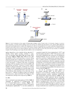
Figure 3. A brief introduction to three popular 3D-bioprinting techniques with cell-laden bioinks. (A) Schematic diagram of extrusion-
based 3D printing. Cell-laden bioinks contained in the micro-syringe are extruded onto the substrate on the collection plate through the
print nozzle by pneumatic pressure or piston. The dimensions of a structure are translated into X, Y, and Z coordinates during printing by a
computer, which controls the nozzle and substrate. (B) Schematic diagram of digital light processing 3D printing. The construct is printed
with the increased height from the photocrosslinkable liquid bioink in the build vat as the build plate moves up vertically. (C) Schematic
diagram of the 3D-printing process using cell-laden bioinks by drop-on-demand inkjet.
applied depending on the materials during deposition . a potential material for tissue engineering in 1997 and
[29]
[7]
Another widely applied printing technique is light- was first applied clinically in 1999 . HA can interact with
[30]
based 3D printing using digital light processing (DLP) cell surface hyaladherins such as Receptor for Hyaluronan
technology (Table 4) (Figure 3B). Different from EBP, Mediated Motility, which is important for cell migration
the printout of DLP is generated from a reservoir filling under the conditions of inflammation and tissue repair .
[31]
with liquid bioink and is attached to the platform above. Thus, it exhibits superior biocompatibility and the ability
As the platform moving up, the height of the 3D construct to promote chondrogenesis. However, implants composed
is then increased . Compared with other 3D-printing of fragments or low-molecular-weight HA lack biological
[24]
techniques, DLP has superior vertical structure fidelity and interaction with encapsulated cells and surrounding tissue,
high printing resolution . However, it usually requires leading to inflammation or degradation of scaffolds. As a
[21]
bioinks to be photocrosslinkable. In addition, the viscosity result, increasing chemical-modified HA derivatives have
of bioinks should be maintained in a specific range so that been developed (Table 2) .
[32]
the printout can withstand dissociation from the bottom HA and its derivatives are widely applied in
of the build vat while attaching to the build plate or the 3D printing, especially EBP . In 2015, Kesti et al.
[38]
layer above . With the exception of these two methods designed a novel bioink by blending polymer poly(N-
[25]
mentioned above, drop-on-demand 3D printing has also isopropylacrylamide) grafted hyaluronan (pNIPAAM)
been applied in cartilage tissue engineering (Table 1). with methacrylated hyaluronan (MeHA) (Table 4). The
high-resolution scaffold generated showed immediate
2.1. HA termination of flow and rapid gelation, but it required the
HA is a polymeric glycosaminoglycan (GAG) (Table 4) elution of HA-pNIPAAM after printing to prevent high
consisting of duplicated β−1,4-d-glucuronic acid- death of embedded cells . Later in 2017, the ultraviolet
[39]
β−1,3-N-acetyl-Dglucosamine residues (Figure 4A). (UV)-crosslinkable MeHA hydrogel was applied to print a
As one of the major components in ECM, HA promotes porous and rigid scaffold. The printout showed improved
chondrogenesis significantly . It was first considered as storage moduli and elastic moduli. However, precise control
[28]
18 International Journal of Bioprinting (2022)–Volume 8, Issue 3