
Page 29 - IJB-8-3
P. 29
Liang, et al.
the superficial layer of the neo-tissue . Stratesteffen also characterized by a low adverse immune reaction,
[61]
et al. developed a GelMA-COL ink via drop-on-demand compatible degradation rates, and superior elasticity .
[66]
3D printing method in 2017 . Using the co-culture B. mori silk without sericin has an initial modulus of
[62]
of human endothelial cells and hMSCs, the addition 15 – 17 Gpa, which is stronger than most other resources.
of COL increased cell spreading, storage modulus, In addition, it can be incorporated with other biopolymers,
and viscosity of this material . In 2018, Yang et al. such as gelatin, to develop particular bioinks, enabling
[62]
assessed the mechanical properties and biocompatibility scaffolds fabricated with tunable mechanical properties
of alginate bioink, alginate/agarose bioink, and alginate/ and controlled pore sizes .
[70]
COL I bioink. Among these three materials, alginate/ The utilization of SF as a natural source of
COL I hydrogel had higher cell viability and cartilage 3D-printing bioinks has advanced rapidly in recent
gene marker expression levels than the other two kinds 3 – 4 years. In 2017, Shi et al. developed a BMSC-
of inks, but with more inferior compressive modulus and laden SF/gelatin bioink for articular cartilage repair .
[71]
tensile strength compared with alginate/agarose one . Scientists observed a significant increase in HYP and
[47]
Simultaneously, another work constructed a 3D-printed GAG accumulation during a 21-day in vitro culture with
porous scaffold via COL crosslinked by tannic acid (TA) the addition of BMSC affinity peptide E7, indicating a
(Table 4), a non-toxic plant polyphenol . This bioink superior chondrogenesis ability. However, the mechanical
[63]
could gelate at a temperature around 37 ℃, suggesting properties of this biomaterial are not shown. In 2019,
that it is applicable in the human body. For its printability, bioink consisting of SF and gelatin was further improved
TA-crosslinked preosteoblast-laden COL bioink was able to be crosslinker-free, as most of the chemical agents
[69]
to be printed into a construct with a pore size of 512 ± added for SF polymerization are toxic . In addition,
46 μm and strut size of 315 ± 10 μm. The findings from a study by Kim et al. developed an advanced SF-based
in vitro experiments also indicated that this COL -based bioink (Sil-MA) by methacrylating SF via glycidyl
construct with an optimal TA concentration of 0.5 wt% methacrylate and built a scaffold for cartilage repair using
[72]
could maintain cell viability of 95% throughout the 14- DLP 3D-printing (Table 4) . Their results showed that
day preosteoclast culture. A recent work conducted by scaffold by bioink composed of 30% Sil-MA exhibited
Wang et al. developed a bi-phasic scaffold with gradient a compressive modulus of 910 kPa, which was able to
mechanical strength via cryogenic 3D printing . TGFβ1- hold a kettlebell weighing 7kg and recovered without any
[64]
loaded COL I hydrogel was filled in the printed frame to deformation after removing the bell. As for its printability,
form the cartilage zone. At 37℃, the compressive strength 30% Sil-MA scaffold with interconnected pores of sized
of the cartilage layer was 0.12 Mpa and the elastic modulus up to 700 μm was successfully printed and the inner
was 1.05 Mpa, which are similar to those of natural human structure was visible to the naked eye. Using 3D-printed
cartilage tissue. Additionally, the shear strength between 30% Sil-MA cartilaginous trachea, significant cartilage
the cartilage zone and the subchondral zone was 0.4 Mpa. matrix formation and the presence of chondrocytes were
The interface also had a peel strength of 470 N/m. These observed after 4 weeks of culture in vitro, suggesting
two results indicated that the ink was capable for cryogenic superior ability to promote cartilage formation and
[72]
3D printing of two-layer osteochondral scaffold. It has also biocompatibility of Sil-MA as a novel bioink .
been demonstrated that the expression of cartilage gene 2.5. GelMA
markers, such as SOX9 and COL II, was significantly
upregulated in the cartilage layer with TGF-β1 through in GelMA is a gelatin derivative that mainly contains
vitro experiments using rat BMSCs . methacrylamide groups with a minority of methacrylate
[64]
groups (Figure 4C). It is usually crosslinked via UV light
2.4. SF illumination with the addition of a photoinitiators, such
SF, mainly produced by Bombyx mori silkworms, is as Irgacure 2925 and lithium acylphosphinate (LAP) salt
composed of 43% glycine, 30% alanine, and 12% serine . (Table 4). The photocrosslinking of GelMA can produce
[65]
Sericin, which is a UV-resistant protein that glues the silk
fibers, needs to be removed by the degumming process to
produce soluble SF . The processed SF is then dissolved
[66]
in solvents such as lithium bromide, formic acid, ionic
liquid, and CaCl2/ethanol/water solvent system .
[67]
Aqueous silk solution can be turned into different forms
and structures, including films/membrane, powder,
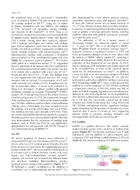
hydrogel, porous sponges, and nanofibers (Figure 5) [68] .
SF hydrogel is usually crosslinked by the addition of
crosslinkers such as glutaraldehyde and genipin . It is Figure 5. Schema of the Bombyx mori silk processing.
[69]
International Journal of Bioprinting (2022)–Volume 8, Issue 3 21