Page 31 - IJB-8-3
P. 31

Liang, et al.                                       PEG  has  been  shown  to  facilitate  chondrogenic  ECM


                                                               regeneration back in 2002 . It is capable for maintaining
                                                                                    [86]
                                                               the viability of non-adhesive cells such as chondrocytes,
                                                               cells including osteoblast and fibroblast . Studies have
                                                                                                [87]
               Full name    Extrusion-based   printing  Digital light processing  Interpenetrating   network  KLF3 Antisense RNA   1: KLF3-AS1  G-protein-coupled   receptor kinase interacting   protein 1: GIT-1   while discouraging the adhesion and spreading of adhesive
                                                               found that this biologically inert property of PEG could
                                                               be  improved  by  the  inclusion  of  hydroxyapatite  and
                                                                                                      . As for its
                                                               Laponite, a kind of synthetic smectite clay
                                                                                                  [88,89]
                                                               mechanical properties, the compressive modulus of PEG
               Acronyms     EBP  DLP    IPN                    hydrogel is about 0.75 MPa, which is relatively stronger
                                                               than other hydrogels but still low compared with that of
                                                               native human articular cartilage .
                                                                                         [90]
                                                                   Scientists have been working on the development of
                                                               PEG hydrogel in 3D printing for the past decade. In 2014,
                  Lithium acylphosphinate salt  Bone marrow-derived   mesenchymal stem cells  Human mesenchymal stem   Transforming growth factor β  of tricalcium phosphate used as bone graft substitute .
                                                               Zhang et al. developed a 3D-printed PEG scaffold with
                                                               β-tricalcium phosphate ceramic, which is a special form
                                                                                                            [91]
                                                               The  scaffold  was  of  50%-65%  porosity  and  was  fully
                                                               interconnected. Formation of new tissue with smooth but
                                                               raised surface was observed 24 weeks after implantation
               Full name                Platelet-rich plasma  Glycosaminoglycan  Hydroxyproline  using  rabbit  model  with  trochlea  defects .  Gao  and
                                                                                                   [90]
                                                               other  scientists  fabricated  a  PEG-GelMA  scaffold
                                   cells
                                                               encapsulating hMSCs by inkjet printing in 2015 . The
                                                                                                        [92]
                                                               contained cells were shown to maintain in their initially
                                                               deposited position during printing and the cell viability
               Acronyms  LAP  BMSC  hMSC  PRP  GAG  TGFβ  HYP  was  over  80%.  63%  of  embedded  hMSCs  underwent
                                                               chondrogenic  differentiation  after  21  days  of  in vitro
                                                               chondrogenesis, but cell hypertrophy was also observed.
                                                               The  compressive  modulus  of  either  cell-laden  or  non-
                                                               cell-laden PEG-GelMA scaffold was also lower than that
                                                               of their corresponding PEG scaffold . In 2018, Wang et
                                                                                             [92]
                                                               and PEG diacrylate (PEGDA) (Table 4) . Their results
                                                                                                 [78]
                                                               showed  that  the  compressive  stress  of  hydrogels  with
               Full name  Polyethylene glycol  Polymer poly   (N-isopropylacrylamide)   grafted hyaluronan  Methacrylated   hyaluronic acid  Polycaprolactone  Methacrylated SF  PEG diacrylate  Tannic acid  al. developed a UV-crosslinkable ink containing GelMA
                                                               both GelMA and PEGDA was significantly higher than
                                                               that of GelMA alone, but the novel ink became fragile as
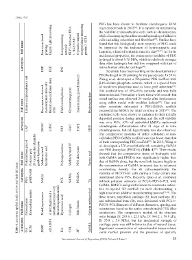
            Table 4. Acronyms applied in this review and their full name
                                                               the concentration of GelMA increased due to enhanced
               Acronyms  PEG  pNIPAAM  MeHA  PCL  Sil-MA  PEGDA  crosslinking  density.  For  its  cytocompatibility,  the
                                                               viability of MC3T3-E1 cells during a 7-day culture was
                                                               maintained above 99%. Recently, Qiao et al. combined
                                                               triblock  polymer  networks  of  PCL-b-PEG-b-PCL  with
                                                         TA
                                                               GelMA, BMSCs and growth factors to construct a native-
                                                               like  tri-layered  3D  scaffold  via  melt  electrowriting,  a
                                                               high-resolution additive manufacturing process
                                                                                                          . The
                                                                                                      [93,94]
                                                               and subchondral bone (B), were fabricated with PCL-b-
               Full name  Osteoarthritis  Extracellular matrix  Metalloproteinase  A disintegrin and a   metalloproteinase with   thrombospondin motifs  Hyaluronic acid  Collagen  Silk fibroin  Gelatin methacryloyl  three layers, superficial cartilage (S), deep cartilage (D),
                                                               PEG-b-PCL filaments of different diameters, spacing, and
                                                               orientations based on the native osteochondral COL fiber
                                                               architecture.  The  compressive  moduli  of  the  structure
                                                               were benign (S: 283.6 ± 22.3 kPa, D: 964.2 ± 56.8 kPa,
                                                               B:  55.8  ±  5.4  MPa),  but  the  mechanical  strength  of
               Acronyms  OA  ECM  MMP  ADAMTS  HA  COL  SF  GelMA  cartilage parts was still inferior to that of natural tissue.
                                                               Significant accumulation of osteochondral tissue-related
                                                               zonal  marker  proteins  and  the  presence  of  spatially

                                       International Journal of Bioprinting (2022)–Volume 8, Issue 3        23
   26   27   28   29   30   31   32   33   34   35   36