
Page 281 - IJB-8-3
P. 281
Zheng, et al.
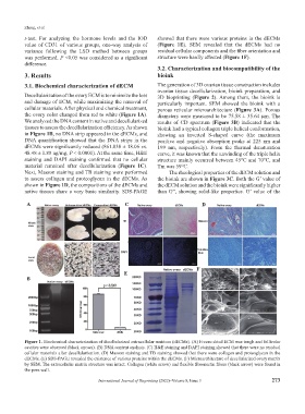
t-test. For analyzing the hormone levels and the IOD showed that there were various proteins in the dECMs
value of CD31 of various groups, one-way analysis of (Figure 1E). SEM revealed that the dECMs had no
variance following the LSD method between groups residual cellular components and the fiber orientation and
was performed. P <0.05 was considered as a significant structure were hardly affected (Figure 1F).
difference.
3.2. Characterization and biocompatibility of the
3. Results bioink
3.1. Biochemical characterization of dECM The generation of 3D ovarian tissue construction includes
ovarian tissue decellularization, bioink preparation, and
Decellularization of the ovary ECM is to minimize the loss 3D bioprinting (Figure 2). Among them, the bioink is
and damage of ECM, while maximizing the removal of particularly important. SEM showed the bioink with a
cellular materials. After physical and chemical treatment, porous reticular microarchitecture (Figure 3A). Porous
the ovary color changed from red to white (Figure 1A). diameters were measured to be 75.58 ± 35.64 µm. The
We analyzed the DNA content in native and decellularized results of CD spectrum (Figure 3B) indicated that the
tissues to assess the decellularization efficiency. As shown bioink had a typical collagen triple helical conformation,
in Figure 1B, no DNA strip appeared in the dECMs, and showing an inverted S-shaped curve (the maximum
DNA quantification showed that the DNA strips in the positive and negative absorption peaks at 225 nm and
dECMs were significantly reduced (861.838 ± 18.06 vs. 199 nm, respectively). From the thermal denaturation
48.48 ± 1.88 ng/mg, P < 0.0001). At the same time, H&E curve, it was known that the unwinding of the triple helix
staining and DAPI staining confirmed that no cellular structure mainly occurred between 45°C and 70°C, and
material remained after decellularization (Figure 1C). Tm was 59°C.
Next, Masson staining and TB staining were performed The rheological properties of the dECM solution and
to assess collagen and proteoglycan in the dECMs. As the bioink are shown in Figure 3C. Both the G’ value of
shown in Figure 1D, the compositions of the dECMs and the dECM solution and the bioink were significantly higher
native tissues share a very basic similarity. SDS-PAGE than G”, showing solid-like properties. G’ value of the
A C D
F
E
B
Figure 1. Biochemical characterization of decellularized extracellular matrices (dECMs). (A) Freeze-dried ECM was tough and follicular
cavities were observed (black arrows). (B) DNA content analysis. (C) H&E staining and DAPI staining showed that there were no residual
cellular materials after decellularization. (D) Masson staining and TB staining showed that there were collagen and proteoglycan in the
dECMs. (E) SDS-PAGE revealed the existence of various proteins within the dECMs. (F) Microarchitecture of decellularized ovary matrix
by SEM. The extracellular matrix structure was intact. Collagen (white arrow) and flexible fibronectin fibers (black arrow) were found in
the pore wall.
International Journal of Bioprinting (2022)–Volume 8, Issue 3 273