
Page 284 - IJB-8-3
P. 284
3D-bioprinted Ovary Initiated Puberty in the Model Mice
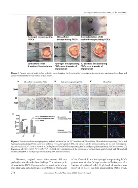
Figure 5. General view of grafts before and after transplantation. At 4 weeks after implantation, the constructs maintained their shape and
developed functional blood vessels (black arrow).
A B C
D E F
H
G
Figure 6. Evaluation of the neoangiogenesis and cell proliferation. (A-C) The effects of 3D scaffolds, 3D scaffold encapsulating POCs, and
hydrogel encapsulating POCs constructs on blood revascularization (CD31, red arrows). (D-F) Immunostaining for the cell proliferation-
specific marker (Ki67, cells in brown), in the implanted 3D scaffold encapsulating POCs and hydrogel encapsulating POCs constructs. (G)
Expression of CD31, Ki67. *P < 0.05, **P < 0.0001. (H) Identification of the apoptosis of sections from grafts treated with 3D scaffold
encapsulating POCs and hydrogel encapsulating POCs (white arrows).
However, vaginal smear observations did not of the 3D scaffold and the hydrogel encapsulating POCs
perfectly coincide with these findings. The estrous cycle groups were similar (a large number of leukocyte and a
of mice in the OVX-C group rested on anestrus for a long fraction of epithelial cells). High level of anestrus was
time that were called estrous cycle inhibition. The results observed in the 3D scaffold encapsulating POCs group,
276 International Journal of Bioprinting (2022)–Volume 8, Issue 3