
Page 80 - IJB-8-3
P. 80
Protein Nanoparticles Promote Cell Growth in 3D Bioprinted Constructs
was then placed on top of a heat-bed set to 40°C to create 2.10. Cytoskeletal staining and imaging
a suitable temperature environment for the cells. Immunostaining was performed at each culture time
Three syringe pumps were set up to dispense
homogenous gel and extrude peptide bioink through point. In brief, the cells were fixed with 4% (v/v)
paraformaldehyde solution for 30 min and incubated in a
the nozzle. The first syringe pump was loaded with cold cytoskeleton buffer (3 mM MgCl , 300 mM sucrose,
peptide solution and set to a flow rate of 55 μL/min. and 0.5% [v/v] Triton X-100 in PBS solution) for 5 min
2
The second pump was loaded with ×7 PBS and set to to permeabilize the cell membranes. The permeabilized
a flow rate of 20 μL/min. The third pump was loaded cells were then incubated in blocking buffer solution (5%
with the cells and set to a flow rate of 20 μL/min. FBS, 0.1% (v/v) Tween-20, and 0.02% [w/v] sodium
Three samples of the 4-layer rectangular prism were azide in PBS) for 30 min at 37°C. For F-Actin, anti-mouse
printed for each condition with a height of 1.5 mm per IgG (whole molecule)-FITC and rhodamine-phalloidin
sample to facilitate imaging. The same procedure was (1:300) were added to the cells for 1 h. Further, the cells
conducted for samples without gas vesicles to serve as were incubated in DAPI for 5 min to counterstain the
controls. nucleus. The fluorescent dye-treated cells were observed
2.8. 3D cell proliferation assay and imaged using a laser scanning confocal microscope
(Zeiss LSM 710 Inverted Confocal).
The CellTiter-Glo® luminescent 3D cell viability assay
was used to assess cell proliferation in 3D hydrogels by 2.11. Scanning electron microscopy (SEM)
measuring ATP production. At each time point, the kit The printed samples were characterized using SEM to
was equilibrated at room temperature for approximately visualize the morphology of the peptide bioink and gas
30 min. CellTiter-Glo® Reagent equal to the volume of vesicles in printed samples . Samples were printed on
[50]
cell culture medium present in each well was added. The 18 × 18 mm glass coverslips and given time to solidify
contents were mixed for 5 min to digest the hydrogels before dehydration by gradual immersion in increasing
and then incubated for 30 min. After incubation, concentrations of 20%, 40%, 60%, 80%, and 100% (v/v)
the luminescence was recorded using a plate reader ethanol solutions for 5 min in each solution. Further
(PHERAstar FS, Germany). dehydration in 100% ethanol solution was done by
2.9. Live/dead staining changing the absolute ethanol solution with a fresh ethanol
solution twice for 5 min each, followed by a third time
HEK293 cells were seeded into peptide according to the for 2 h. Dehydrated samples were subsequently placed
protocol described above. After one, three, and 7 days into the critical point dryer (Sorvall Critical Point Drying
of incubation, the media was removed and replaced System) for evaporation before being mounted onto SEM
with PBS containing approximately 2 mM calcein AM aluminum pin stubs with double-stick conductive carbon
and 4 mM ethidium homodimer-1 before incubation tape and a final sputter coating of 10 nm of iridium.
for 40 min. Before imaging, the staining solution was Images were taken with FEI Magellan XHR SEM. This
removed, and fresh PBS was added. Stained cells were was done with a TLD detector, and imaging parameters
imaged under an inverted confocal microscope (Zeiss included a current of 50 pA, a high voltage of 3.00 kV, and
LSM 710 Inverted Confocal Microscope, Germany). The a working distance of 4.0 m. Biological peptide hydrogel
percentage of living cells was obtained through analysis coating cells were fixed with 2.5% (v/v) glutaraldehyde
with ImageJ. (diluted from 25%) in water overnight at 4°C, the post-
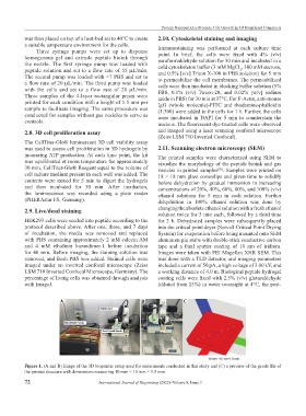
A B C
Figure 1. (A and B) Image of the 3D bioprinter setup used for experiments conducted in this study and (C) a preview of the gcode file of
the printed structure with dimensions measuring 10 mm × 10 mm × 1.5 mm.
72 International Journal of Bioprinting (2022)–Volume 8, Issue 3