
Page 82 - IJB-8-3
P. 82
Protein Nanoparticles Promote Cell Growth in 3D Bioprinted Constructs
A B C D
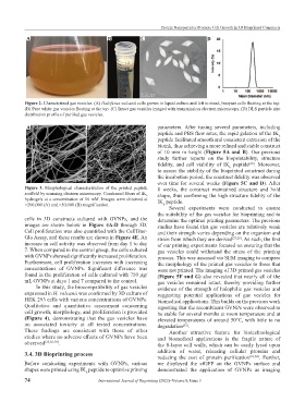
Figure 2. Characterized gas vesicles. (A) Haloferax volcanii cells grown in liquid culture and left to stand, buoyant cells floating at the top.
(B) Pure white gas vesicles floating at the top. (C) Intact gas vesicles imaged with transmission electron microscopy. (D) DLS particle size
distribution profile of purified gas vesicles.
A B parameters. After tuning several parameters, including
peptide and PBS flow rates, the rapid gelation of the IK
6
peptide facilitated smooth and consistent extrusion of the
bioink, thus achieving a more refined and stable construct
of 10 mm in height (Figure 5A and B). Our previous
study further reports on the bioprintability, structure
fidelity, and cell viability of IK peptide . Moreover,
[60]
6
to assure the stability of the bioprinted construct during
the incubation period, the construct fidelity was observed
over time for several weeks (Figure 5C and D). After
Figure 3. Morphological characterization of the printed peptide 8 weeks, the construct maintained structure and hold
scaffold by scanning electron microscopy. Condensed fibers of IK shape, thus confirming the high structure fidelity of the
6
hydrogels at a concentration of 16 mM. Images were obtained at
×200,000 (A) and ×50,000 (B) magnification. IK peptide.
6
Several experiments were conducted to ensure
the suitability of the gas vesicles for bioprinting and to
cells in 3D constructs cultured with GVNPs, and the determine the optimal printing parameters. The previous
images are shown below in Figure 4A‑D through 3D. studies have found that gas vesicles are relatively weak
Cell proliferation was also quantified with the CellTiter- and their strength varies depending on the organism and
Glo Assay, and these results are shown in Figure 4E. An strain from which they are derived [27,29] . As such, the first
increase in cell activity was observed from day 1 to day of our printing experiments focused on ensuring that the
7. When compared to the control group, the cells cultured gas vesicles could withstand the stress of the printing
with GVNPs showed significantly increased proliferation. process. This was assessed via SEM imaging to compare
Furthermore, cell proliferation increases with increasing the morphology of the printed gas vesicles to those that
concentrations of GVNPs. Significant difference was were not printed. The imaging of 3D printed gas vesicles
found in the proliferation of cells cultured with 750 μg/ (Figure 5F and G) also revealed that nearly all of the
mL GVNPs at days 1 and 7 compared to the control. gas vesicles remained intact, thereby providing further
In this study, the biocompatibility of gas vesicles evidence of the strength of halophilic gas vesicles and
expressed in H. volcanii was confirmed by 3D culture of suggesting potential applications of gas vesicles for
HEK 293 cells with various concentrations of GVNPs. biomedical applications. This builds on the previous work
Qualitative and quantitative assessment concerning reporting that the recombinant GVNPs were observed to
cell growth, morphology, and proliferation is provided be stable for several months at room temperature and at
(Figure 4), demonstrating that the gas vesicles have elevated temperatures of around 50°C, with little to no
no associated toxicity at all tested concentrations. degradation .
[35]
These findings are consistent with those of other Another attractive feature for biotechnological
studies where no adverse effects of GVNPs have been and biomedical applications is the fragile nature of
observed [35,56-59] . the S-layer cell walls, which can be easily lysed upon
3.4. 3D Bioprinting process addition of water, releasing cellular proteins and
. Further,
reducing the cost of protein purification
[61,62]
Before conducting experiments with GVNPs, various we displayed the sfGFP on the GVNPs surface and
shapes were printed using IK peptide to optimize printing demonstrated the application of GVNPs as imaging
6
74 International Journal of Bioprinting (2022)–Volume 8, Issue 3