
Page 138 - IJB-8-4
P. 138
3D Arenas for C. elegans Behavior
in z dimension (plane perpendicular to the assay plate made of 2% agar-based NGM hydrogel. The resulting
surface) is essentially limited to varying depth of the arenas are nematode-friendly, minimizing the stress that
arenas. Vertical elements, suspended features, multilevel could have been induced when the animals are transferred
structures, and other similar architectures are not allowed from the culture plate into the arenas.
using molds. To demonstrate the suitability of the Parnon-printed
To meet the need for alternative fabrication processes, parts, we used them to assess C. elegans physical barrier
we explored two methods. The first one includes the use of crossing ability, in the context of aging (young, middle-
polyvinyl alcohol (PVA), a water-soluble synthetic polymer, aged adults), feeding history (fully fed [FF], starved
to cast NGM structures (Figure 1). The ensuing parts are of animals), and prior experience (have been or not in the
high quality and provide valuable feedback regarding the presence of a 3D structure before). We also explored
NGM self-sustaining properties. However, this method’s the usage of 3D-printed structures to spatially confine
limitations motivated us to seek another route, one that C. elegans egg laying behavior. C. elegans behavior
employs a 3D printer, which uses NGM as ink. in 3D environments is by definition not possible to be
A growing number of researchers are employing explored on standard flat NGM plates. Therefore, the
3D printing as a transformative tool for cell and tissue findings reported here would likely not have been brought
engineering [14,15] . This includes 3D scaffolds made of to light if the Parnon Printer had not been developed.
enriched hydrogel-based materials . Hydrogels of 1 –
[16]
5% agar concentrations have been successfully explored 2. Methods
for 3D bioprinting applications . Most of the occurring
[17]
structures are sturdily self-supported cubes or other In this section, we describe the PVA casting and the
non-hollow, no-overhang designs. Interestingly, the Parnon printing methods, and C. elegans behavioral
3D-printing technology has not been used to produce experiments process. Technical details on the printer’s
behavioral arenas for the study of small invertebrate customization, NGM rheological properties, and software
animal models, like C. elegans. communication between the printer’s parts can be found
We present a highly customized prototype 3D in the Supplementary File. A list of the major components
printer, the Parnon Printer (Parnon: Mountain in South used for the conversion of the commercial printer into
Greece, known for its many gorges), which can print 3D Parnon is provided in the Supplementary File. Details on
parts, suitable for C. elegans behavioral experiments, the Arduino code are provided in the Supplementary File.
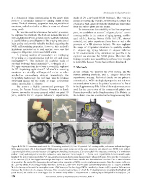
A B E
C D F
Figure 1. NGM 3D structures made with the PVA casting method. (A) Two 3D-printed PVA casting molds, red arrows indicate the liquid
NGM pouring input. (B) A four-legged NGM crossbridge, made using one of the casts shown in (A), placed on a NGM plate surface.
Legs are slightly tilted outwards because of flipping and handling the structure. (C) A diving bell-like structure, consisting of a hemisphere
(radius: 5 mm) and designed to have five cylindrical arms, diameter: 3 mm, length: 1 – 1.5 mm, each. Liquid NGM did not reach the entire
length of the hollow space inside the PVA cast, resulting in much shorter arms than originally planned (4 mm). Note also the rough surface
of the structure. (D) A four-legged crossbridge standing on a 2 mm thick base, raised 4 mm above the base’s surface. This is a much thinner
structure than the one in (B), showcasing the self-supporting properties of NGM even in smaller arrangements. Note the missing right arm.
(E) Close-up of the NGM diving bell-like structure, shown in C, side view. Yellow frame indicates the position of a C. elegans nematode.
Note the bumpy surface and the incomplete beams. (F) Close-up of the four-legged NGM crossbridge, shown in (B), top view. Yellow frame
indicates the position of a C. elegans nematode. Note the very rough surface, of which the protruding features (examples highlighted with
green arrows) are similar to or even bigger than the worm’s body width. C. elegans worm is challenging to distinguish in both (E) and (F).
130 International Journal of Bioprinting (2022)–Volume 8, Issue 4