
Page 164 - IJB-8-4
P. 164
Recombinant Human Collagen for 3D Bioprinting of Skin Equivalent
had functional basal HaCaTs expressing proliferative 3.5. In vivo evaluation of bioinks
marker K14 at week 3, while the differentiation marker K10
was seldom observed. This is probably because the in vitro To further explore the feasibilities of using composite
skin constructs were at an early stage of differentiation bioinks containing rhCol3 to the in vivo skin repair and
under ALI condition (3 weeks since the ALI culture). In regeneration, we developed a full-thickness skin defect
contrast, differentiation marker K10 was significantly model of SD rats. The GelMA-rhCol3-3.2 and GelMA
detected at week 6 in all groups, indicating that the were selected to treat the skin wounds by depositing
in vitro skin construct formed a more differentiated upper hydrogel precursors on the wounded site followed by
epidermal layer. The fluorescent intensity of the marker in situ photocrosslinking. The skin tissue repair and
K14 and K10 was quantified and is shown in Figure 5D. regeneration were evaluated by measuring wound closure
There were no significant differences in the intensity rate and by examining the histological staining.
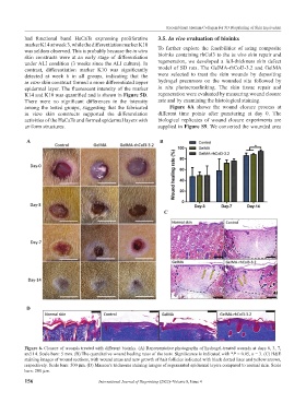
among the tested groups, suggesting that the fabricated Figure 6A shows the wound closure process at
in vitro skin constructs supported the differentiation different time points after puncturing at day 0. The
activities of the HaCaTs and formed epidermal layers with biological replicates of wound closure experiments are
uniform structures. supplied in Figure S9. We converted the wounded area
A B
C
D
Figure 6. Closure of wounds treated with different bioinks. (A) Representative photographs of hydrogel-treated wounds at days 0, 3, 7,
and 14. Scale bars: 5 mm. (B) The quantitative wound healing rates of the tests. Significance is indicated with *P < 0.05, n = 3. (C) H&E
staining images of wound sections, with wound areas and new growth of hair follicles indicated with black dotted lines and yellow arrows,
respectively. Scale bars: 500 μm. (D) Masson’s trichrome staining images of regenerated epidermal layers compared to normal skin. Scale
bars: 200 μm.
156 International Journal of Bioprinting (2022)–Volume 8, Issue 4