
Page 305 - IJB-8-4
P. 305
Gu, et al.
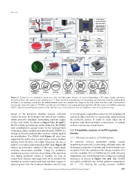
Figure 2. Fabrication of a perfusable vessel-on-a-chip. (A) Fabrication process: (i) coaxial bioprinting of EC-laden tubular constructs;
(ii) cutting fibers into segments and culturing for 5–7 days; (iii) PCL stent printing; (iv) demolding and sterilizing stents by soaking them
in ethanol; (v) inserting a stent into the endothelialized vessel as a support (the image on the left, which was taken with a fluorescence
microscope, shows the status of HUVECs growth and proliferation); (vi) casting the hydrogel bulk with the vessel and GelMA containing
VEGF; and (vii) assembling the perfusion chip. (B) Top view of the perfusion chip. (C) Explosive view of the perfusion chip.
design of the perfusion chamber ensured sufficient of several quickly-separable connectors, this modularized
contact between the hydrogel bulk and culture medium, perfusion chip could also be conveniently separated from
which provided abundant surrounding nutrient supply the perfusion system. It could be easily taken out of
for the cells inside. As shown in Figure 2Avi, B, and C, incubator and observed under a microscope, if needed,
the PLA frame and adequate cavity limited by the PDMS without introducing contamination.
wall contributed to enough room for the hydrogel bulk
contacting culture medium omnidirectionally. PDMS is a 3.2. Printability analysis of GelMA/gelatin
biological friendly polymer that has been widely applied bioinks
in microfluidics. The PDMS wall Figure 2C used here 3.2.1. Rheological analysis of GelMA/gelatin
supported the entire chip and prevented culture medium
leaking when the bolts were tight. Moreover, the use of To optimize printing parameters during coaxial
hollow cover plates and transparent PET films Figure 2B bioprinting for precisely constructing cell-laden tubes, the
offered an observable window of the chip, which made rheological properties of gelatin and GelMA bioink were
real-time observation possible. Using an incubation measured. First, the variation of the storage modulus (G′)
monitoring system, real-time observation of the sprouting and loss modulus (G″) of GelMA/gelatin with changing
area during perfusion culture is feasible. As long as temperature was tested during both cooling and warming
proper focal distance and range were set in advance, the processes, as shown in Figure 3Ai and Aii. GelMA
monitoring system would send back real-time images of and gelatin exhibited very similar gelation temperatures
sprouting area from the incubator. Besides, with the help (G′ = G″). During the cooling process, GelMA/
International Journal of Bioprinting (2022)–Volume 8, Issue 4 297