
Page 308 - IJB-8-4
P. 308
Vessel-on-a-Chip for Antiangiogenic Drug Screening
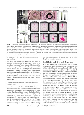
Figure 4. Comparison of hydrogel constructs with/without a PCL stent. (A) Mechanical properties of the hydrogel bulk with/without a
stent. (i) Photo of the hydrogel bulk with a stent compression test. (ii) Stress-strain curves of the hydrogel bulks with/without a stent. (iii)
Stresses of the hydrogel bulk with/without a stent at a strain of 50% and 60%. (B) Patency of the hydrogel tubes with/without a stent after
swelling. Dashed circle represents the inner wall of the channel; scale bar: 500 μm. (i) Optical image of the hydrogel tube without a stent
after swelling (axial direction). (ii) Optical image of the hydrogel tube with a stent after swelling (axial direction). (iii) Inner diameters of
the hydrogel tubes with/without a stent after swelling. (C) Photos of the hydrogel tubes with/without a stent perfusion performance test at
flow rates of 6 μl/min, 60 μl/min, 600 μl/min, and 6000 μl/min.
3.3.2. Patency of hydrogel tubes with/without a stent showed that a PCL stent served was a key factor in the
after swelling long-term perfusion process.
Not only the mechanical properties, but also the 3.4. Diffusion analysis of the hydrogel tube
swelling characteristics of hydrogels need to be
considered during perfusion. In general, hydrogel tubes For the purpose of characterizing the diffusional
reach swelling equilibrium after being immersed in permeability of hydrogel materials and the barrier function
culture medium for 24 h, leading to the expansion of of the endothelialized fabricated vessel, FITC labeled
the tube wall and the narrowing of the inner channel. dextran with a molecular weight of 40 kDa was utilized.
After reaching swelling equilibrium, the introduction As illustrated in Figure 5G, after injecting FITC-dextran
of a PCL stent guaranteed that the inner diameter of solution into the hydrogel tube, fluorescence microscopy
the hydrogel tube would be 1212 μm, while the one images were captured at 5–80 min, at intervals of
without a stent possessed a channel diameter of 879 μm 15 min. Hydrogel tube without cells (Figure 5A-C)
and endothelialized hydrogel tube, which was cultured
(Figure 4B).
7 days after printing (Figure 5D-F), were tested under
3.3.3. Perfusion performance of hydrogel tubes with/ the same condition. In terms of general trends, dextran
without a stent was gradually diffused from the central channels to the
surrounding hydrogel tube (Figure 5A and D); the gray
As stated above, GelMA (EFL-GM-30) is a soft level distribution (Figure 5B and E) became broader
hydrogel that may lead to tube collapse during perfusion. and more dispersed with time, and the curve of which
A perfusion performance test was conducted to assess was flattened; moreover, as illustrated in the gray level
whether liquid could flow through the hydrogel tubes distribution histograms in Figure 5C and F, the gray
with/without a stent at different flow rates (Figure 4C, levels mainly concentrated on high values at the beginning
Videoclip S1). Four flow rates from 6 μl/min to of the test, and the peak of the gray levels moved toward
6000 μl/min were selected. At all flow rates, liquid could lower values as time passed. Obviously, the diffusion
flow through smoothly with the help of the PCL stent, rates in HUVEC-laden tube were much slower than that
while the tube without a stent faced the problem of wall in the cell-free tube. Even in the beginning of the test,
collapse resulting in liquid backflow. These findings without the barrier function of endothelial cells, diffusion
300 International Journal of Bioprinting (2022)–Volume 8, Issue 4