
Page 310 - IJB-8-4
P. 310
Vessel-on-a-Chip for Antiangiogenic Drug Screening
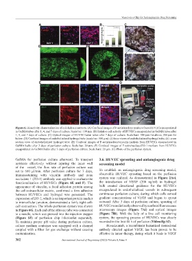
Figure 6. Bioactivity characterization of cell-laden constructs. (A) Confocal images of F-actin/nucleus markers from HUVECs encapsulated
in GelMA tubes after 1, 4, and 7 days of culture. Scale bar: 100 μm. (B) Relative cell activity of HUVECs encapsulated in GelMA tubes after
1, 4, and 7 days of culture. (C) Optical images of HUVEC-laden tubes after 7 days of culture. Scale bars: 500 μm for above, 200 μm for
below. (D) Confocal images of endothelialized hydrogel tube (scale bar: 500 μm): (i) three views of endothelialized hydrogel tube; (ii) cross
section view of endothelialized hydrogel tube. (E) Confocal images of F-actin/nucleus/vinculin markers from HUVECs encapsulated in
GelMA bulks after 3 days of perfusion culture. Scale bar: 20 μm. (F) Confocal images of F-actin/nucleus/ZO-1 markers from HUVECs
encapsulated in GelMA bulks after 3 days of perfusion culture. Scale bars: 20 μm. (G) Photo of the perfusion system.
GelMA for perfusion culture afterward. To transport 3.6. HUVEC sprouting and antiangiogenic drug
nutrients effectively without injuring the inner wall screening model
of the vessel, the flow rate of perfusion culture was
set to 600 μl/min. After perfusion culture for 3 days, To establish an antiangiogenic drug screening model,
immunostaining with vinculin antibody and zona observable HUVEC sprouting based on the perfusion
occludens 1 (ZO-1) antibody was applied to evaluate the system was realized. As demonstrated in Figure 2Avi,
functionalization of HUVECs (Figure 6E and F). The the introduction of VEGF (200 ng/ml) in hydrogel
appearance of vinculin, a focal adhesion protein among bulk created directional guidance for the HUVECs
the cell-extracellular matrix, confirmed a firm adhesion encapsulated in endothelialized vessels in subsequent
between HUVECs and hydrogel was generated. The continuous perfusion culture, during which cells sensed
expression of ZO-1, which is an important protein marker gradient concentrations of VEGF and began to sprout
to intercellular junction, demonstrated a fairly tight cell- outward. After 3 days of perfusion culture, spouting of
cell connection. The whole perfusion system was shown HUVECs was distinctly observed by confocal fluorescence
in Figure 6G. Each end of the silicone tube was connected microscopy images (Figure 7Ai) and optical images
to a needle, which was pierced into the injection stopper (Figure 7Bi). With the help of a live cell monitoring
(Figure S5) of perfusion chip inlet/outlet separately. system, the sprouting process of HUVECs was clearly
To maintain proper pH value during perfusion culture, recorded in the first 48 h of perfusion (Videoclip S2).
culture medium container was equipped with a channel Bevacizumab, a recombinant humanized monoclonal
coupled with a filter for gas exchange without causing antibody directed against VEGF, has been proven to be
contamination. effective in tumor therapy, during which it binds to VEGF
302 International Journal of Bioprinting (2022)–Volume 8, Issue 4