
Page 36 - IJB-8-4
P. 36
Xie, et al.
the printed breast tumor tissue, which indicated the great
potential of this bioink system in various biomedical
applications.
Funding
This study was sponsored by the National Key Research
and Development Program of China (2018YFA0703000,
Yong He), the National Natural Science Foundation
of China (No. U1909218, Yong He), the Science Fund
for Creative Research Groups of the National Natural
Science Foundation of China (No. T2121004, Yong He).
Acknowledgments
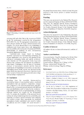
Figure 9. Bioprinting of centimeter-scale breast tumor tissue with
angiogenesis. This study was sponsored by the National Key Research
and Development Program of China (2018YFA0703000,
converge with each other. Due to the increasing of VEGF Yong He), the National Natural Science Foundation
in the 3D environment excreted by the encapsulated of China (No. U1909218, Yong He), the Science Fund
MDA-MB-231s, the 3D angiogenesis phenomenon was for Creative Research Groups of the National Natural
more obvious and the 3D vessel network became more Science Foundation of China (No. T2121004, Yong He).
complex. The results showed that in vitro bioprinting of
centimeter-scale breast tumor tissue with angiogenesis Conflict of interest
was successfully achieved based on the proposed TSM-B, All authors declare no financial/commercial conflicts of
demonstrating its capability in more corresponding interest.
biomedical applications in future.
Centimeter-scale tissues based on hydrogel Author contributions
materials constructed by 3D bioprinting technology are
of great application value. However, the question of the Conceptualization: Yong He, Jianzhong Fu, Zichen Chen
substances exchanging inside and outside centimeter- Investigation: Mingjun Xie, Yuan Sun, Zhenliang Fu
scale structure and 3D angiogenesis inside large-scale Methodology: Mingjun Xie
structure has restricted the development of centimeter- Formal analysis: Mingjun Xie, Ji Wang, Lei Pan
scale tissues. Combining to the hot spot of “secondary Writing - original draft: Mingjun Xie
printing” of microspheres currently and the bran-new Writing - review & editing: Yong He, Ji Wang
requirements of bioprinting to researchers, an innovative
microsphere-based bioink system was invented in this References
paper, which was expected to become an important model
in the research of tumor developing and anti-cancer drugs 1. He Y, Gu Z, Xie M, et al., 2020, Why Choose 3D Bioprinting?
corresponding to angiogenesis. Part II: Methods and Bioprinters. BioDesign Manuf, 3:1–4.
https://doi.org/10.1007/s42242-020-00064-w
4. Conclusion 2. Thakor J, Ahadian S, Niakan A, et al., 2020, Engineered
Benefiting from the reversible thermo-sensitive Hydrogels for Brain Tumor Culture and Therapy. BioDesign
crosslinking feature of non-modified gelatin, sacrificial Manuf, 3:203–26.
microspheres that can respond to temperature changes https://doi.org/10.1007/s42242-020-00084-6
were prepared by the electrohydrodynamics principle. 3. Lee M, Bae K, Guillon P, et al., 2018, Exploitation of
Based on this, on-demand nutrient channels with different
sizes and distribution could be formed in the centimeter- Cationic Silica Nanoparticles for Bioprinting of Large-Scale
scale GelMA structures by sol-gel transferring, providing Constructs with High Printing Fidelity. ACS Appl Mater
sufficient nutrient and oxygen for the encapsulated Interfaces, 10:37820–8.
cells. To examine the feasibility of the proposed bioink https://doi.org/10.1021/acsami.8b13166
system, the sol-gel transferring behavior, printability, and 4. Ying GL, Jiang N, Maharjan S, et al., 2018, Aqueous Two-
capability of nutrient channel formation were analyzed
and verified in detail. More importantly, the sacrificial Phase Emulsion Bioink-Enabled 3D Bioprinting of Porous
microspheres loaded endothelial cells could be further Hydrogels. Adv Mater, 30:1805460.
vascularized and form complex 3D vessel network in https://doi.org/10.1002/adma.201805460
28 International Journal of Bioprinting (2022)–Volume 8, Issue 4