
Page 34 - IJB-8-4
P. 34
Xie, et al.
A B
C
D E
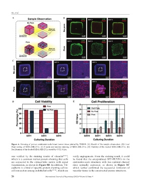
Figure 6. Growing of porous centimeter-scale breast tumor tissue printed by TSM-B. (A) Sketch of the sample observation. (B) Live/
Dead testing of MDA-MB-231s. (C) F-actin and nucleus staining of MDA-MB-231s. (D) Viability of the loaded MDA-MB-231s. (E)
Proliferation of the loaded MDA-MB-231s tested by CCK-8 kits.
was verified by the staining results of vinculin [39-41] , verify angiogenesis. From the staining result, it could
which is a common marker protein showing that cells be found that the encapsulated GFP-HUVECs in the
are connected to the extracellular matrix with signal centimeter-scale structures with two nutrient channel
transmission, as shown in Figure 8B. In addition, VE- sizes normally expressed, as shown in Figure 8C
cadherin is a kind of specific protein showing cell-to- which further confirmed the successful formation of
cell connection among endothelial cells [42-44] , which can vascular tissue in the constructed porous structures.
26 International Journal of Bioprinting (2022)–Volume 8, Issue 4