
Page 35 - IJB-8-4
P. 35
Microsphere-Based Bioink for Large Tissue with Angiogenesis
A B
C
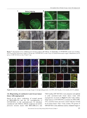
Figure 7. Angiogenesis in the centimeter-scale structures printed with TSM-B. (A) Distribution of GFP-HUVECs at the start of culture.
th
(B) Fluorescence microscope images of 3D sprout of GFP-HUVECs on the 5 day. (C) Optical microscope images of attachment and 3D
nd
rd
sprout of GFP-HUVECs on the 2 and 3 day.
A C
B D
Figure 8. Confocal fluorescence microscope images of corresponding proteins. (A) CD31, (B) Vinculin, (C) β-tubulin, (D) VE-cadherin.
3.8. Bioprinting of centimeter-scale breast tumor TSM loading GFP-HUVECs were prepared and applied
tissue with angiogenesis to print centimeter-scale breast tumor tissue with
angiogenesis. As shown in Figure 9, after 7-day culture,
Based on the above verification of normal growth
of MDA-MB-231s and the 3D vascularization of longer sprout of GFP-HUVECs toward to the MDA-MB-
GFP-HUVECs in the centimeter-scale 3D structures 231s could be found and more vessel branches formed
constructed with TSM-B, TSM-B composed of GelMA in the tumor tissue. After 12-day culture, 3D sprouts of
precursor solution mixed with MDA-MB-231s and GFP-HUVECs from different nutrient channels began to
International Journal of Bioprinting (2022)–Volume 8, Issue 4 27