
Page 45 - IJB-8-4
P. 45
Lu, et al.
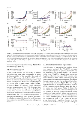
A B C
D E F
G H
I
Figure 2. Compressive mechanical properties of PVA/dECM hydrogels. (A) Uniaxial stress-strain curves under compression until 80% of
height. (B) Uniaxial compression-relaxation curves of PVA/dECM hydrogel group and control group. The compression-relaxation cycles of
the same gel for 20 consecutive cycles without any time interval between two cycles, including control (C), 20-20 (D), 40-20 (E), and 120-
20 (F) PVA/dECM hydrogel groups. (G) The normalized maximum compressive stress of the hydrogels in the 20 cycles. (H) The dissipation
energy of the hydrogels in the 20 cycles. (I) The stress-relaxation of the hydrogels. The strain of each loading step increased by 10% in the
range of 0 – 50%.
recover the original shape after folding (Figure 3C) 3.5. Evaluation of meniscus regeneration
and stretching (Figure 3D). After 12 weeks of implantation, the meniscus defects
3.4. Biocompatibility test were repaired eminently by PVA/dECM hydrogels.
Immunological rejection or synovial proliferation was
HUVECs were cultured on the surface of various not observed in any of these groups. The defective
hydrogels at the same initial concentration to assess region of the PVA/dECM hydrogel group was covered
the biocompatibility of the materials. The results of by glossy and smooth meniscus, which was close to the
the Calcein/PI staining and phalloidine/DAPI staining natural meniscus. In the control and blank groups, the
showed an obvious adhesion morphology and a large defects were filled with regenerated tissues, which were
number of living cells in the PVA/dECM hydrogel group irregular, depressive, and clearly distinguishable from
and control group (Figure 4 and Figure S15-S16). the surrounding meniscus (Figure 5A). The ImageJ
As shown in Figure S17, although the cell viability of software showed that the regeneration area of the PVA/
hydrogel groups decreased after 48 h of culture, there dECM meniscus was significantly larger than that in
are still a large number of living cells on the hydrogel the control and blank groups (Table 1). Meanwhile,
the contact surface between femur and tibial plateau in
surface. Alkali treatment and freeze-thaw cycle had little the PVA/dECM hydrogel group was smooth without
effect on cell compatibility. Overall, the above-mentioned roughness. Obvious cartilage defects were observed in
results revealed that PVA/dECM hydrogel group had a the contact surface between femur and tibial plateau in
promising biocompatibility, and it would be appropriate the control and blank groups. The macroscopic scores
for cell adhesion and proliferation, which could be used as are presented in Table 1. The PVA/dECM hydrogel
tissue engineering material for constructing and repairing group demonstrated a significant increase in all the
meniscus. assessments. In the ICRS scoring system, the cartilage in
International Journal of Bioprinting (2022)–Volume 8, Issue 4 37