
Page 48 - IJB-8-4
P. 48
A strong bio-ink for Meniscus Regeneration
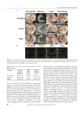
A
B
Figure 5. (A) The general view of the regenerated meniscus and worn cartilage surface. The blue dotted line showed the regeneration of
meniscus and the wear of cartilage on the surface of femoral and tibial plateau. (B) The micro-MRI of the rabbit knee after 12 weeks. The
red arrow indicates the regeneration site of meniscus defect.
Table 1. The area of meniscal repair and the total scores of ICRS properties may be made using this technique. It was also
and O’Driscoll. discovered that when water was added to the created PVA
Group Area of Total Total hydrogel, 90% of the plastic deformation due to extension
meniscal ICRS O’Driscoll was recovered, resulting in a significant contraction force
[17]
repair (%) score score capable of lifting items 1100 times their weight . The
PVA/dECM 92.37±3.68 10±1.08 20.33±2.15 PVA/dECM hydrogel showed a promising compression
Control 60.94±6.52 7±1.16 16.33±1.89 performance, and it was not broken when compressed to
Blank 45.23±7.32 6±1.50 14.66±2.67 80%. The Young’s modulus reached 0.49 MPa, and the
stress limitation was 2.9 MPa. An excellent compression
ability allows hydrogels to keep intact in meniscal repair.
have been shown to be a key regulator of proper tissue
shape when used in physiological settings. Meanwhile, The mechanical environment has a big impact on cellular
functions such cell adhesion, cell tension, and cytoskeleton
multiple in vivo and in vitro investigations have revealed architecture, and it even has a long-term stability
that mechanical variables play a key role in the meniscus’s influence. According to Zhang et al., cyclic hydrostatic
health, degeneration, and regeneration [32,33] . The PVA/ compress force therapy stimulates proliferation of isolated
dECM hydrogel scaffold showed a promising mechanical primary meniscus fibrochondrocytes through increasing
strength. The PVA forms stable crystalline domains by a integrin 51 expression . An appropriate mechanical
[34]
high concentration of hydroxyl ion and repeated freezing/ environment plays an important role in the regeneration
thawing cycles. Physically crosslinked PVA biomaterials and repair of meniscus. Meanwhile, excellent mechanical
with excellent mechanical capabilities, low water properties are conducive to the protection of cartilage,
content, damage resistance, and shape memory polymer which reduced friction between cartilage surfaces.
40 International Journal of Bioprinting (2022)–Volume 8, Issue 4