
Page 49 - IJB-8-4
P. 49
Lu, et al.
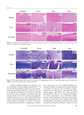
Figure 6. Histological results of the H&E staining (×200 magnification; scale bar: 50 μm). Histology and gross morphology at upper right
corner (×40 magnification; scale bar: 500 μm).
Figure 7. Histological results of the toluidine blue staining (×200 magnification; scale bar: 50 μm). Histology and gross morphology at
upper right corner (×40 magnification; scale bar: 500 μm).
A photocrosslinked technique was utilized to 3D and a fast recovery for the PVA/dECM hydrogel. An
print the mixture of sodium alginate and PEGDA [35,36] . It excellent elasticity can simulate the biological functions
can not only form hydrogel quickly, but also possesses of meniscus and play the role of buffering pressure.
satisfactory mechanical properties, which can be used Furthermore, we discovered in this study that adding
as an effective supplement to PVA network. PEGDA sodium alginate and PEGDA to bio-ink resulted in
and bioactive glass nanoparticles containing copper and high printability. Zhang et al. suggested bioenergetics
sodium alginate were used to create a nanocomposite and bone regeneration using 3D-printed double-
scaffold, according to Li et al. The scaffold exhibited the network alginate hydrogels containing polyphosphate.
great biomimetic elastomeric mechanical properties, with The pre-gel combining sodium alginate and PEGDA
a high compressive strength of 6.1 kPa . The design exhibited higher 3D printing performance than typical
[35]
of a double network provides a high-pressure resistance hydrogels for manufacturing complex scaffolds for tissue
International Journal of Bioprinting (2022)–Volume 8, Issue 4 41