
Page 46 - IJB-8-4
P. 46
A strong bio-ink for Meniscus Regeneration
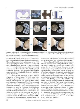
A B
C
D
Figure 3. 3D printing test. (A) Schematic diagram of upper meniscus model of 3D printer and the printing hydrogel meniscus. Uniform
holes can be observed under a microscope. (B) Meniscus model size on the computer and the actual size. (C) Folding of the printed meniscus
and recovery. (D) Stretching of the printed meniscus and recovery.
the PVA/dECM hydrogel group showed a minor damage homogeneous in the PVA/dECM group, which indicated
and the same height level with the surrounding cartilage. that the meniscus has been well regenerated (Figure 9).
The overall assessment was roughly normal for the PVA/ The cartilage in PVA/dECM group showed a smooth
dECM hydrogel group (mean score was 10 ± 1.08), and structure similar to natural cartilage from H&E staining.
abnormal for the control and blank groups (mean scores The results of staining with toluidine blue, Safranin O, and
were 7 ± 1.16 and 6 ± 1.50, respectively). The well filled collage Ⅱ in the PVA/dECM group showed the cartilage
meniscus was observed in the PVA/dECM hydrogel matrix strong staining and the uniform cell distribution.
group using micro-MRI, and the detective and displaced Immature chondrocytes arranged parallel to the surface
meniscus could be visualized in the control and blank of cartilage and mature chondrocytes were distributed
groups (Figure 5B). in groups in cartilage lacunae, which was similar to
According to the results of the H&E staining natural cartilage. These results showed the well cartilage
(Figure 6), the defects in the PVA/dECM group were protection in the PVA/dECM hydrogel group. Compared
filled by the regenerated tissue that was intact and uniform. with the PVA/dECM group, the color of ECM in the
Obvious gap and defects were observed in the control control and blank groups was dimmed, and an obvious
and blank groups. The meniscus ECM of the regenerated gap could be observed. The arrangement of immature
region showed a strong staining with toluidine blue and chondrocytes had no orderly pattern, and the number of
Safranin O in the PVA/dECM group, indicating the active cartilage lacunae was reduced. In the O’Driscoll scoring
regeneration of meniscus. The cells in the defected area system, the cellular morphology, Safranin O staining,
retained the morphology and were interweaved in the structural integrity, cartilage thickness, and cellularity
PVA/dECM group, which was similar to that in the natural were used to evaluate the cartilage conditions and the PVA/
meniscus. However, no defection healing was found in dECM hydrogel group showed the best performance. The
the control and blank groups, in which irregularly shaped mean scores for the PVA/dECM hydrogel, control, and
cells were found in the defected area (Figures 7 and 8). blank groups were 20.33 ± 2.15, 16.33 ± 1.89, and 14.66
The staining of collage Ⅱ on ECM was pronounced and ± 2.67, respectively (Table 1).
38 International Journal of Bioprinting (2022)–Volume 8, Issue 4