
Page 50 - IJB-8-4
P. 50
A strong bio-ink for Meniscus Regeneration
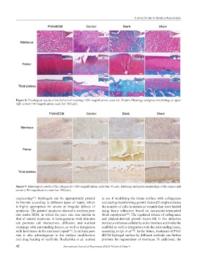
Figure 8. Histological results of the Safranin O staining (×200 magnification; scale bar: 50 μm). Histology and gross morphology at upper
right corner (×40 magnification; scale bar: 500 μm).
Figure 9. Histological results of the collagen II (×200 magnification; scale bar: 50 μm). Histology and gross morphology at the upper right
corner (×40 magnification; scale bar: 500 μm).
engineering . Hydrogels can be appropriately printed to see if modifying the tissue surface with collagenase
[37]
by bio-ink according to different types of injury, which and adding transforming growth factor-β3 might enhance
is highly appropriate for severe or irregular defects of the number of cells in meniscus wounds that were healed
meniscus. The printed meniscus showed a uniform pore using tissue adhesives based on isocyanate-terminated
size under SEM, in which the pore size was similar to block copolymers . The regulated release of collagenase
[39]
that of natural meniscus. A homogeneous void structure and platelet-derived growth factor-AB in the defective
can promote cell interactions, diffusion, and nutrient meniscus enhances cellularity at the interface and inside the
exchange with surrounding tissues, as well as integration scaffold, as well as integration with the surrounding tissue,
with host tissue in the meniscal repair . A uniform pore according to Qu et al. . In the future, treatment of PVA/
[38]
[40]
size is also advantageous to the surface modification dECM hydrogel surface by different methods can further
and drug loading of scaffolds. Bochyńska et al. wanted promote the regeneration of meniscus. In additional, the
42 International Journal of Bioprinting (2022)–Volume 8, Issue 4