
Page 92 - IJB-8-4
P. 92
3D Bioprinting of Human Neural Tissues
A
B
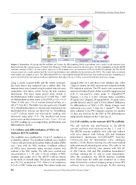
Figure 1. Preparation of tunicate dECM scaffolds and bioinks for differentiating NSCs to peripheral nerve tissues. Fresh tunicates were
harvested from the coastal regions of United Arab Emirates (UAE) and processed in the laboratory. (A) The preparation of sterile dECM
scaffolds, seeding of NSCs, in vitro culture, induction of PN differentiation, and formation of PN on the dECM scaffolds. (B) The dECM
was processed into a sterile powder and the bioink was formulated and optimized by incorporating the NSCs. The neural tissue constructs
were printed and cultured in vitro. The differentiated PN was characterized after the PN induction. Our results proved that bioprinting is a
promising method for translational medicine applications that support tissue viability, tissue differentiation, and tissue storage.
using a sterile surgical knife and the whole hydrogel- changed after 24 h and then every alternate day. After
like tunic tissue was separated into a culture dish. The 3 days of culture, the NSC medium was replaced with the
tunicate tissue was cleaned using deionized water at room PN induction medium. The PN media was composed of
temperature few times, before being cut into required neurobasal media (Thermo fisher scientific) supplemented
dimensions. The tunic tissue pieces were stirred in with 1× non-essential amino acids, 1× GlutaMAX™
decellularization buffer consisting of 10 mM Tris, 1 mM (Sigma), 1 X N2, 1 X B27 (Thermo fisher scientific),
of ethylenediamine tetra acetic acid (EDTA), 0.2% v/v of 20 ng/ml EGF (Sigma), 20 ng/ml bFGF, 10 ng/ml nerve
Triton X-100, and 1.5% of sodium dodecyl sulfate, at a growth factor-β, and 25 µM Y27632 (Merck Millipore)
pH of 7.5 for 48 h. The buffer was changed every 2 h until for differentiation of NSCs to PN. Media changes were
10 h. Detailed description on the tunicate material and the performed once every 3 days for 2 weeks . The cells
[24]
decellularization process are described by Govindharaj were cultured in the PN induction medium for another
et al. Cellular debris was removed by washing with 12 days and checked for the NSC to PN differentiation
[23]
deionized water after 10 h. The decellularized tissue using specific markers at day 3 and day 12.
pieces were cut into dimensions of 1 cm × 1 cm × ~0.1 cm
for NSC seeding for cytocompatibility, proliferation, and 2.4. Cell viability in the tunicate dECM scaffolds
differentiation studies. The cell viability and proliferation in the dECM
2.3. Culture and differentiation of NSCs on tunicate scaffolds were analyzed on day 3, 7, and 12.
tunicate dECM scaffolds The dECM tunicate scaffolds with cells and without
cells were stained with Calcein AM and Ethidium
The scaffolds were sterilized for 1 h in UV irradiation in homodimer1 (Invitrogen LIVE/DEAD™ Viability/
a biosafety cabinet. The sterilized scaffolds were washed Cytotoxicity Kit, for Mammalian Cells, catalogue
3 times with prewarmed phosphate-buffered saline (PBS) number L3224). Before staining, the cells were washed
and 1 time with the NSC medium. Confluent cultures with prewarmed physiological saline. The cells in the
of NSCs were harvested using accutase enzyme and dECM tunicate scaffolds were stained with 500 µL
washed in the NSC media. The cells were counted and of 2M Calcein AM and 4M Ethidium homodimer1
concentrated to 3 × 10 cells in 30 µL volume of NSC working solution for 45 min at room temperature.
6
medium and seeded on to the dECM scaffolds placed After the incubation, the dECM tunicate scaffolds
in the wells of 24-well plate. The plates were incubated were lifted from the wells and mounted on a clean
in a 5% CO incubator at 37°C. After 4 h of incubation, slide followed by confocal imaging using a Leica SP8
2
0.5 mL of NSC media was added. The culture media was confocal laser scanning microscope. In this staining
84 International Journal of Bioprinting (2022)–Volume 8, Issue 4