
Page 95 - IJB-8-4
P. 95
Soman, et al.
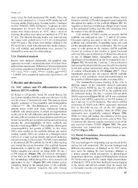
twice using the fresh prewarmed PN media. Then, the clear morphology of peripheral neuronal fibers, which
tissues were immersed in 1:1 ratio of PN media and cell formed a network of PN with elongated neural outgrowth
freezing media (Embryomax freezing media, Catalogue throughout the surface of the scaffolds (Figure 2F). We
number ES002D, EMD Millipore), incubated at room hypothesize that these morphology changes might be due
temperature for 10 min. The 24-well plate containing to the mechanical surface cues the cells experience from
tissues were frozen directly at −80°C. After 1 week of the surface of the dECM scaffold.
freezing, the plates were taken out and kept at 37°C for Cell viability of NSCs seeded on tunicate dECM
15 min. The thawed freezing media was removed and scaffolds was analyzed on days 3, 7, and 12 of culture.
the tissues were washed twice in 0.5 mL of prewarmed Live-dead staining was used to see the viable cells on
fresh PN media. The tissues were further cultured in the scaffolds and colorimetric AlamarBlue assay was used
PN media for a week with alternate day media changes. for the quantification of cell proliferation. The live-dead
The cell viability and proliferation were assessed by assay of cells grown on the tunicate dECM scaffolds
AlamarBlue assay and live-dead staining. showed an increase in the number of green florescent
cells over time and proliferation of thread-like structures
2.16. Statistical analysis by day 12 (Figure 2J-L). AlamarBlue assay showed
Results were analyzed statistically. All graphical data significant cell proliferation on day 12 compared to day 3
represent the mean ± standard deviation of at least three (Figure 2N). Between day 3 and day 7, the proliferation
independent experiments. Differences between treatments was not significant, probably because the cells are adapting
were tested using the two-tailed Student’s t-test. *P < 0.05, to the new culture environment along with the pressure
[27]
**P < 0.01, ***P < 0.001, ****P < 0.0001, and *****P of cellular differentiation to PN (Figure 2N) . These
< 0.00001 were considered statistically significant in all experiments proved that the tunicate dECM scaffolds
cases. provide a very conducive tissue microenvironment for
the growth of different types of cells, including NSCs.
3. Results and discussion The immunofluorescence staining of the cells in
different stages of the induction showed expression
3.1. NSC culture and PN differentiation in the of PN marker, NEFH by day 12, which indicated the
tunicate dECM scaffolds differentiation of the NSCs to PN on the scaffolds with
To design a cell-specific bioink for neural tissues, the the induction media. The day 3 controls did not show
key factors considered in this study were the type of cells NEFH expression (Figure 2G-I). qPCR was performed
and the choice of biomaterials used. The tunicate tissue to analyze the mRNA expression of the PN markers on
majorly composed of biocompatible and biodegradable day 3 and day 12 of PN induction. The relative mRNA
cellulose [23,26] . The decellularization process of the expression of NEFH and PRPH expression increased
tunicate tissue could yield clean, transparent looking four to five-fold on day 12 of the PN induction,
scaffolds with natural pores, in which we hypothesized compared to the day 3 samples, while the HNK1 gene
would aid in the cell adherence and proliferation. Since expression was remarkably reduced (Figure 2M).
the stem cells are sensitive to many intrinsic and extrinsic HNKI is a stem cell marker, the reduction of this gene
factors for growth such as matrix coating and cell seeding indicates the differentiation of stem cells to PNs. Day
density, the cytocompatibility of these scaffolds was 12 neurons also showed slightly increased levels of the
initially tested using MEFs . The scaffolds offered good pan neuronal marker TUBB3 (Figure 2M). The gene
[23]
cytocompatibility and growth of MEFs. In this work, expression was normalized to house-keeping gene
NSCs were seeded on the scaffolds without any matrix GAPDH (Figure 2M).
coating (usually NSCs are seeded on a Matrigel-coated 3.2. Bioink formulation, characterization, cross-
surface) at a high seeding density and neural induction linking, and bioprinting
was given on day 3 of the culture. The NSCs were attached
to the tunicate dECM scaffolds by 24–48 h after seeding The base hydrogel for bioprinting was optimized before
and started the colony formation. The scaffolds became adding the cell component. The tunicate hydrogel and
impermeable to light by day 3 (Figure 2A-C). Hence, different concentrations of Matrigel in the NSC media
further imaging was carried out using SEM. On day 3 were used for making the base hydrogel. Matrigel is
of PN induction (Figure 2D), the electron micrograph a matrix protein which polymerizes at physiological
of the NSCs on the scaffolds still showed the fibroblast- temperature; therefore, aliquots of Matrigel were stored at
like morphology, which changed to a more rounded cell 4°C before mixing with NSC media. High concentrations
appearance by day 7 (Figure 2E) with distinct colony of Matrigel (>30%) in the base hydrogel caused clogging
formation, filling in the natural pores of the scaffolds. of the needle and did not extrude from the nozzle due
By day 12 of neuronal induction, the cells showed to increased viscosity and rapid solidification within the
International Journal of Bioprinting (2022)–Volume 8, Issue 4 87