
Page 100 - IJB-8-4
P. 100
3D Bioprinting of Human Neural Tissues
A B C
D E F
G H I
J K L
M
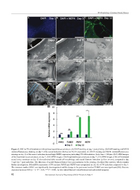
Figure 5. NSC to PN differentiation in bioprinted neural tissue constructs. (A) DAPI staining on day 1 post-printing. (B) DAPI staining and NEFH
immunofluorescence staining on day 3 of the neural induction showed no NEFH expression. (C) DAPI staining and NEFH immunofluorescence
staining on day 12 of the neural induction showed high NEFH expression, indicating PN differentiation. Scale bars = 100 µm. (D-F) SEM images
of the bioprinted tissue constructs on day 3. (G-I) SEM images of the bioprinted tissue constructs on day 7. (J-L) SEM images of the differentiated
neural tissue construct on day 12 showed remarkable neural cell morphology and neural filament formation (yellow arrows), compared to day
3 and day 7 post-induction. The direction of neural filament formed were perpendicular to the printing direction (blue arrows), which requires
further investigation. (M) mRNA expression of PN markers PRPH and NEFH were upregulated on day 12 of PN induction compared to day 3.
The stemness marker HNK1 was significantly downregulated and the change in the pan neural marker TUBB3 was non-significant (NS). Data are
expressed as mean+SD (n = 3; *P < 0.05, ***P < 0.001, by two- tailed Student’s t-test between test and control samples).
92 International Journal of Bioprinting (2022)–Volume 8, Issue 4