
Page 101 - IJB-8-4
P. 101
Soman, et al.
A C method efficiently uses an untapped source of biomaterial
to formulate tissue-specific bioinks. The development of
sustainable bioinks from marine invasive tunicates would
open up new avenues for scaling up the hydrogel-based soft-
tissue bioprinting for translational medicine applications.
Acknowledgment
B D This research was partially carried out using the Core
Technology Platforms resources at New York University
Abu Dhabi.
Funding
The research was funded by the NYUAD startup grant for
Sanjairaj Vijayavenkataraman.
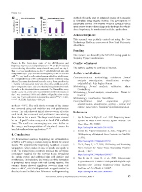
Figure 6. The freeze-thaw study of the dECM-grown and Conflict of interest
bioprinted neurons. (A) Cell proliferation of PN on dECM scaffold
evaluated using AlamarBlue assay showed less growth on day 3, The authors declare no conflicts of interest.
then recovered proliferation on day 7, but still showed less cells
compared to day 1. (B) Live-dead staining of day 7 dECM scaffold Author contributions
with PN; very less live cells noticed compared to bioprinted tissues.
(C) Cell proliferation of PN on bioprinted tissues evaluated using Conceptualization, methodology, validation, formal
AlamarBlue assay also showed less cells on day 3 compared to day analysis, investigation, visualization, writing–
1, but the cells recovered faster and showed two-fold growth by original draft: Soja Saghar Soman
day 7 compared to day 1. (D) Live-dead staining also showed more Methodology, formal analysis, validation: Mano
live cells in the bioprinted tissue constructs. For AlamarBlue assay Govindharaj
results (A and C), viable cells recovered from the frozen tissues on Methodology, formal analysis, visualization: Noura Al
day 1 was considered 100% and relative cell proliferation on day Hashimi
3 and day 7 were calculated in AlamarBlue assay (***P < 0.001, Methodology, visualization: Jiarui Zhou
****P < 0.0001). Scale bars = 500 µm.
Conceptualization, fund acquisition, project
administration, visualization, writing – review and
media at −80°C. The cold shock recovery of the tissues editing, supervision: Sanjairaj Vijayavenkataraman
was assessed using cell viability and cell proliferation
assays. The cells showed initial slow recovery after the References
cold shock, but recovered and proliferated on culturing
them further for a week. The bioprinted tissues showed 1. Qiu B, Bessler N, Figler K, et al., 2020, Bioprinting Neural
better cell proliferation compared to the dECM scaffold- Systems to Model Central Nervous System Diseases. Adv
tissue. The results are encouraging to explore further on Funct Mater, 30:1910250.
the storage and transportation of bioprinted tissues for https://doi.org/10.1002/adfm.201910250.
translational medicine applications [37,38] .
2. Soman SS, Vijayavenkataraman S, 2020, Perspectives on
4. Conclusions 3D Bioprinting of Peripheral Nerve Conduits. Int J Mol Sci,
We demonstrated seamless bioprinting and differentiation 21:5792.
of NSCs to PN using a custom-designed bioink for neural https://doi.org/10.3390/ijms21165792
tissues. We optimized the bioprinting workflow at room 3. Yu X, Zhang T, Li Y, 2020, 3D Printing and Bioprinting
temperature, which makes it easy to handle and quick to Nerve Conduits for Neural Tissue Engineering. Polymers
print. The printed tissue constructs maintain the soft-tissue (Basel), 12:1637.
consistency required for the nervous tissue throughout https://doi.org/10.3390/polym12081637
the culture period and exhibited high cell viability and 4. Gao F, Xu Z, Liang Q, et al., 2019, Osteochondral
proliferation. On induction, the bioink aided the formation
of peripheral nerve tissues with well-formed neurites. The Regeneration with 3D-Printed Biodegradable High-Strength
cultured tissues showed significant recovery from cold Supramolecular Polymer Reinforced-Gelatin Hydrogel
shock at −80°C, which holds promise in using this method to Scaffolds. Adv Sci (Weinh), 6:1900867.
develop tissues for clinical use. Moreover, our bioprocessing https://doi.org/10.1002/advs.201900867
International Journal of Bioprinting (2022)–Volume 8, Issue 4 93