
Page 97 - IJB-8-4
P. 97
Soman, et al.
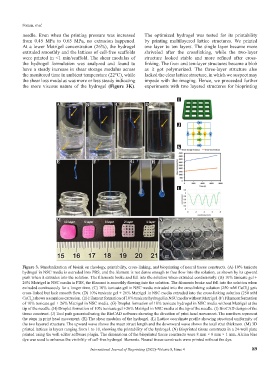
needle. Even when the printing pressure was increased The optimized hydrogel was tested for its printability
from 0.45 MPa to 0.65 MPa, no extrusion happened. by printing multilayered lattice structures. We printed
At a lower Matrigel concentration (26%), the hydrogel one layer to ten layers. The single layer became more
extruded smoothly and the lattices of cell-free scaffolds shriveled after the crosslinking, while the two-layer
were printed in <1 min/scaffold. The shear modulus of structure looked stable and more refined after cross-
the hydrogel formulation was analyzed and found to linking. The five- and ten-layer structures became a blob
have a steady increase in shear storage modulus across as it got polymerized. The three-layer structure also
the monitored time in ambient temperature (22°C), while lacked the clear lattice structure, in which we suspect may
the shear loss modulus was more or less steady indicating impede with the imaging. Hence, we proceeded further
the more viscous nature of the hydrogel (Figure 3K). experiments with two layered structures for bioprinting
I
A B C D
J
K
E F G H
L
M N
Figure 3. Standardization of bioink on rheology, printability, cross-linking, and bioprinting of neural tissue constructs. (A) 10% tunicate
hydrogel in NSC media is extruded into PBS, and the filament is not dense enough to free flow into the solution, as shown by its upward
push when it extrudes into the solution. The filaments broke and fell into the solution when extruded continuously. (B) 10% tunicate gel +
26% Matrigel in NSC media in PBS, the filament is smoothly flowing into the solution. The filaments broke and fell into the solution when
extruded continuously for a longer time. (C) 10% tunicate gel in NSC media extruded into the crosslinking solution (250 mM CaCl ) gets
2
cross-linked but lack smooth flow. (D) 10% tunicate gel + 26% Matrigel in NSC media extruded into the cross-linking solution (250 mM
CaCl ) shows a seamless extrusion. (E) Filament formation of 10% tunicate hydrogel in NSC media without Matrigel. (F) Filament formation
2
of 10% tunicate gel + 26% Matrigel in NSC media. (G) Droplet formation of 10% tunicate hydrogel in NSC media without Matrigel at the
tip of the needle. (H) Droplet formation of 10% tunicate gel +26% Matrigel in NSC media at the tip of the needle. (I) BioCAD design of the
tissue construct. (J) Tool path generated using the BioCAD software showing the direction of print head movement. The numbers represent
the steps in print head movement. (K) The shear modulus of the hydrogel. (L) Lattice coordinate profile showing structural uniformity of
the two layered structure. The upward wave shows the mean struct length and the downward wave shows the total strut thickness. (M) 3D
printed lattices in layers ranging from 1 to 10, showing the printability of the hydrogel. (N) Bioprinted tissue constructs in a 24-well plate
printed using the well editor software plugin. The dimensions of the bioprinted tissue constructs were 8 mm × 8 mm × 1 mm. Alcian blue
dye was used to enhance the visibility of cell-free hydrogel filaments. Neural tissue constructs were printed without the dye.
International Journal of Bioprinting (2022)–Volume 8, Issue 4 89