
Page 230 - IJB-9-1
P. 230
International Journal of Bioprinting Cellulose-based bio-inks for bone and cartilage TE
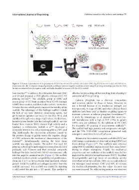
Figure 5. Schematic representation of the preparation of GCA bio-ink and 3D scaffolds. (A) Gelatin-CMC-Alg (GCA) bio-ink loaded with MG 63 os-
teosarcoma cells. (B) A computer-designed polylactic acid-based meniscus negative mold was printed using 3D printing technology, and then the GCA
bio-ink was extruded into the negative mold, and finally demolded to become a 3D-Bio-GCA scaffold.
base reaction to enhance the interaction between CMC effective for preventing cell loss resulting from shearing in
[80]
and CS and prepared a CMC-glycolic chitosan (GC) 3D conventional 3D bioprinting.
printing bio-ink . The aldehyde group of CMC and Calcium phosphate has a chemical composition
[81]
amine group of GC form an imine bond (C=N) through and structure similar to those of bones. However, its
Schiff’s base reaction, and there is also an ionic interaction use is limited because of its mechanical strength and
between the two, which greatly improves the stability of the microporosity. A range of CMC and other cellulose-based
scaffold. The advantages of this hydrogel scaffold include additives have been used as binders or gelling agents to
the lack of any toxic chemical crosslinking agents, fast maintain cohesion in calcium phosphate formulations .
[84]
gel formation (gelation can occur in less than 40 s), and A study by Montelongo et al. showed that novel bio-
stability of the gel over a range of pH values. Furthermore, ink formulations with a high β-TCP (75%) to gelatin
lactoferrin was loaded into the hydrogel scaffold, and the (25%) ratio are stabilized by the addition of 3% CMC
loaded bone marrow MSCs showed high activity and a for successful 3D printing of macroporous scaffolds .
[85]
tendency to differentiate. Sathish et al. developed a The scaffold had high porosity and compressive strength,
[82]
composite trimeric bio-ink comprising gelatin, CMC, and and the 75% TCP+CMC composition promoted early
Alg. Additionally, the electrostatic attraction caused by osteogenic commitment and cell adhesion.
the positive charge of gelatin versus the negative charge
of CMC enhances the mechanical characteristics of the Mohan et al. fabricated a composite scaffold of NFC and
material . Simultaneously, creating the bone tissue CMC using a combination of direct ink writing (DIW) and
[83]
scaffold was equally creative. The author first used 3D freeze-drying (Figure 6) . The scaffolds were obtained
[86]
printing technology to make negative meniscal molds from using DIW to establish large pores and then using freeze-
polylactic acid filaments and then extruded composite drying to obtain micropores. Finally, the mechanical
hydrogel bio-inks loaded with MG63-osteosarcoma cells properties and wet resilience of the scaffold were further
into the negative meniscal molds (Figure 5). The scaffold improved via dehydrothermal treatment (DHT). Excellent
exhibited excellent cellular compatibility and proliferation. biocompatibility and high levels of osteoblastic activity
From day 1 to 7, as the incubation time increased, cellular were observed on the scaffolds. DHT can not only enhance
growth inside the scaffold and collagen secretion from the the hydrogen bond between CMC and NFC to increase
scaffold increased. This scaffold was created by injecting the mechanical properties, but also adjust the pore size
a cell-loaded bio-ink into a 3D model. This method is via water removal. No cellulose degradation was observed
V 222 https://doi.org/10.18063/ijb.v9i1.637
Volume 9 Issue 1 (2023)olume 9 Issue 1 (2023)