
Page 364 - IJB-9-1
P. 364
International Journal of Bioprinting Micro/nano-3D hemostats for rapid wound healing
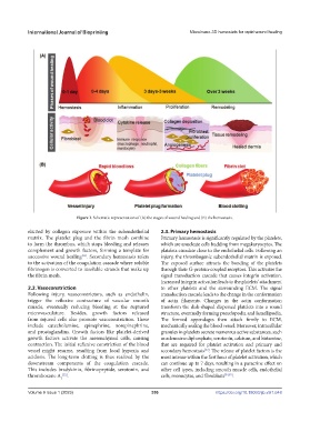
Figure 1. Schematic representation of (A) the stages of wound healing and (B) the hemostasis.
elicited by collagen exposure within the subendothelial 2.3. Primary hemostasis
matrix. The platelet plug and the fibrin mesh combine Primary hemostasis is significantly regulated by the platelets,
to form the thrombus, which stops bleeding and releases which are anucleate cells budding from megakaryocytes. The
complement and growth factors, forming a template for platelets circulate close to the endothelial cells. Following an
successive wound healing . Secondary hemostasis refers injury, the thrombogenic subendothelial matrix is exposed.
[31]
to the activation of the coagulation cascade where soluble The exposed surface attracts the bonding of the platelets
fibrinogen is converted to insoluble strands that make up through their G-protein-coupled receptors. This activates the
the fibrin mesh. signal transduction cascade that causes integrin activation.
Increased integrin activation leads to the platelets’ attachment
2.2. Vasoconstriction to other platelets and the surrounding ECM. The signal
Following injury, vasoconstrictors, such as endothelin, transduction cascade leads to the change in the conformation
trigger the reflexive contracture of vascular smooth of actin filaments. Changes in the actin conformation
muscle, eventually reducing bleeding at the ruptured transform the disk-shaped dispersed platelets into a round
microvasculature. Besides, growth factors released structure, eventually forming pseudopodia and lamellipodia.
from injured cells also promote vasoconstriction. These The formed appendages then attach firmly to ECM,
include catecholamine, epinephrine, norepinephrine, mechanically sealing the blood vessel. Moreover, intracellular
and prostaglandins. Growth factors like platelet-derived granules in platelets secrete numerous active substances, such
growth factors activate the mesenchymal cells, causing as adenosine diphosphate, serotonin, calcium, and histamine,
contraction. The initial reflexive constriction of the blood that are required for platelet activation and primary and
vessel might resume, resulting from local hypoxia and secondary hemostasis . The release of platelet factors is the
[33]
acidosis. The long-term clotting is thus realized by the most intense within the first hour of platelet activation, which
downstream components of the coagulation cascade. can continue up to 7 days, resulting in a paracrine effect on
This includes bradykinin, fibrinopeptide, serotonin, and other cell types, including smooth muscle cells, endothelial
thromboxane A [32] . cells, monocytes, and fibroblasts [34,35] .
2
Volume 9 Issue 1 (2023)olume 9 Issue 1 (2023) 356 https://doi.org/10.18063/ijb.v9i1.648
V