
Page 366 - IJB-9-1
P. 366
International Journal of Bioprinting Micro/nano-3D hemostats for rapid wound healing
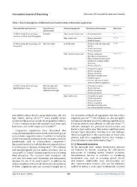
Table 1. Structural properties of fabricated micro/nanostructures in hemostatic applications
Hemostat fabrication process Type of micro/ Structural properties Mechanism of hemostasis References
nanostructure
3D/4D printing, electrospinning, Nanotube High structural aspect ratio • Plasma interaction [155]
extrusion methods, stereolithography
High surface area • Plasma interaction [39,61,156]
• Fluid adsorption
3D/4D printing, electrospinning, soft Micro/nanofiber Small diameter • Physical mesh-like hemostatic [157-160]
lithography barrier
• Platelet interaction
• Plasma interaction
High structural aspect ratio • Platelet entrapment [7,60,90,161-164]
• Red blood cell entrapment
• Blood component entrapment
• Mechanical strength inhibits
blood loss
• Platelet adhesion
High surface area • Drug delivery agent [7,37,161-163,165-167]
• Platelet adsorption
• Plasma adsorption
• Red blood cell adsorption
• Fluid adsorption
• Intrinsic pathway activation
promotes plasma coagulation
3D/4D printing, electrospinning, Micro/nanoparticle, Small size • Drug delivery agent [140,168-171]
digital light processing Micro/nanosphere, • Tissue adhesion
Adhesive powder/gel • Platelet adsorption
• Plasma adsorption
High surface area • Drug delivery agent [131,172-175]
• Platelet adsorption
• Plasma adsorption
• Plasma coagulation
• Red blood cell adsorption
• Ion-induced platelet activation
user-defined release kinetics using temperature, pH, and the promotion of blood cell aggregation that this surface
light stimuli, among others [47,49] . Some popular release roughness permits [17,53] . Microspheres can also strengthen
systems for this purpose include the programmed delivery the hemostat’s intrinsic properties, allowing rapid blood clot
of one or multiple compounds necessary to promote rapid formation and enhanced adhesion to cells and tissue [54-56] .
hemostasis, such as fibrinogen and thrombin . Adhesive microgels are powdered microstructures and
[50]
feature a high surface area. They induce rapid hemostasis
Coagulation experiments have determined that
composite microparticles can both reduce the bleeding time through liquid absorption, forming an in situ hydrogel.
and accelerate coagulation rates, in addition to having the These microgels lead to the concentration of blood cells,
ability to be used in combination with hemostatic adhesive platelets, and other coagulation factors, thus creating a
[57]
powders/gels, such as chitosan-based composites . physical barrier to stop further hemorrhage .
[41]
Microparticles inlaid in scaffolds allow for targeted delivery 3.1.2. Nanoscale structures
of erythrocytes to decrease clotting time . Microspheres At the nanoscale level, surface biochemistry instructs
[51]
with morphologically relevant surface structures, such as cell behavior by directly mediating the cell–material
“macropits” or “craters,” enhance hemostasis by promoting interactions or initiating surface receptor activation.
fluid absorption. The increased fluid absorption rate Nanostructures function as mediators in modulating
and ratio both incur rapid hemostasis and subsequently biochemical cues, such as activating growth factors
decrease the size of the wound area . In addition, the and proteins. Additionally, these nanostructures can
[52]
surface roughness of microscale structures is proportional alter communication between cells and the fabricated
to faster coagulation rates and increased strength due to scaffold to induce a more rapid biochemically activated
V
Volume 9 Issue 1 (2023)olume 9 Issue 1 (2023) 358 https://doi.org/10.18063/ijb.v9i1.648