
Page 376 - IJB-9-1
P. 376
International Journal of Bioprinting Micro/nano-3D hemostats for rapid wound healing
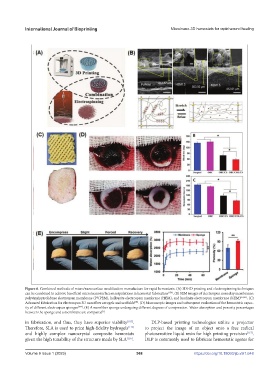
Figure 6. Combined methods of micro/nanosurface modification manufacture for rapid hemostasis. (A) 3D/4D printing and electrospinning techniques
can be combined to achieve beneficial micro/nanosurface manipulations in hemostat fabrication [154] . (B) SEM images of electrospun nanoclay membranes:
polyvinylpyrrolidone electrospun membrane (PVPEM), halloysite electrospun membrane (HEM), and kaolinite electrospun membrane (KEM) [14,41] . (C)
Advanced fabrication for electrospun 3D nanofiber aerogels and scaffolds . (D) Macroscopic images and subsequent evaluation of the hemostatic capac-
[94]
ity of different electrospun sponges [107] . (E) A nanofiber sponge undergoing different degrees of compression. Water absorption and porosity percentages
between the sponge and a membrane are compared .
[7]
in fabrication, and thus, they have superior viability [105] . DLP-based printing technologies utilize a projector
Therefore, SLA is used to print high-fidelity hydrogels [115] to project the image of an object onto a free radical
and highly complex nanocrystal composite hemostats photosensitive liquid resin for high printing precision [117] .
given the high tunability of the structure made by SLA [116] . DLP is commonly used to fabricate hemostatic agents for
V 368 https://doi.org/10.18063/ijb.v9i1.648
Volume 9 Issue 1 (2023)olume 9 Issue 1 (2023)