
Page 372 - IJB-9-1
P. 372
International Journal of Bioprinting Micro/nano-3D hemostats for rapid wound healing
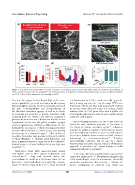
Figure 4. Electrospun micro/nanosurfaces for rapid hemostasis. (A) Scanning electron microscopy (SEM) image of microfibrin fibers adhered on
[37]
[83]
CNFs-coated cotton fibers after clot shrinkage . (B) Blood loss of hemostatic gauzes . (C) Effect of nanofiber weight on mass and the time of bleed-
[91]
ing [152] . (D) Effects of fiber diameter on mechanical properties .
structure, the sponge has low density, high surface area, the development of a CNF-coated cotton fiber gauze for
and compressibility potential, and feature an ultra-strong facile dressing removal after clot shrinkage. CNFs were
liquid absorption capacity. In vitro assessments confirmed transferred onto the clot after facile detachment, resulting
the good cytocompatibility and biodegradability of in smooth cotton fiber and a hairy clot surface. Results
the fabricated hemostatic sponge, as well as its ability indicated that the CNF gauze helps form a gel-like clot
to accelerate the formation of platelet embolism while that properly seals wounds, while an open wound remains
activating both the intrinsic and extrinsic coagulation under any control gauze.
pathways in its mechanism of hemostasis. Further in vivo
assessments demonstrated the gelatin nanofiber sponge’s Electrospinning techniques are also widely used and
ability to rapidly induce stabilization of blood clots, thereby valued for their submicron precision in fabrication in
[67]
leading to the least amount of blood loss compared to other the design of various hemostatic agents . Submicron
commercialized products currently in use, thus proving precision in design is necessary because tunable factors,
its advantage as a hemostatic agent . Other studies on such as morphology, mechanical, electrical, and magnetic
[7]
nanofiber composites have also demonstrated the ability stimulations, influence the type and extent of cell–substrate
[74]
of these structures to promote rapid hemostasis and interactions in micro/nanostructures . Therefore,
incur minimal secondary damage due to the composites’ electrospinning techniques can be used as a micro/
intrinsic property of super-hydrophobicity and high user nanostructure fabrication method, which is a biomimetic
tunability [37,83] . approach to hemostasis and wound healing to inhibit
undesirable side effects optimally .
[45]
Electrospun fibers allow nanoscopic-level control
over factors, such as fiber surface modifications and To this end, CNFs are used extensively because of their
architecture [84,85] . Along with material choice, this conductive properties [14,86-88] . Furthermore, incorporating
control allows for tunability of the desired effect, such as CNFs into hydrogels allows for a biomimetic, electrically
hemostatic capacity and adherence strength. For example, conductive environment that provides a platform for
Figure 4A shows a study by Li et al. , which described further development of controlled drug release and
[37]
Volume 9 Issue 1 (2023)olume 9 Issue 1 (2023)
V 364 https://doi.org/10.18063/ijb.v9i1.648