
Page 187 - IJB-9-2
P. 187
International Journal of Bioprinting A regulated GelMA-MSCs scaffold by three-dimensional bioprinting
The rabbits used in the experiments were provided by To quantitatively detect the degree of osteogenic
the Animal Experiment Center of Soochow University, differentiation and chondrogenic differentiation of MSCs
and all animal experiments were approved by the after induction, four specific genes, namely, BMP-2, Runx-
Experimental Animal Ethics Committee of the First 2, Collagen-1, and SOX9, were selected for detection. Trizol
Affiliated Hospital of Soochow University. The approval was used to extract RNA from MSCs, and in the real-time
ID is SUDA20220530A01. The audit was carried out under reverse-transcription polymerase chain reaction (RT-PCR)
the supervision of the corresponding supervision and experiment, extracted RNA was reverse-transcribed into
management agency. 5 mL of air was slowly injected into cDNA. 2× SYBR Green Mix, Bulge-LoopTM Forward
the ear vein to cause air embolism so as to euthanize an Primer, and Bulge-LoopTM Reverse Primer (RiboBio,
animal. After being soaked in 75% ethanol, the lower limbs China) were added, which were, then, topped up with
were amputated, the hair was removed, and the muscles ddH O to 20 μL. The mixture was put into a quantitative
2
were removed in a sterile environment by soaking in PBS PCR thermal cycler for amplification (Applied Biosystems,
containing 1% double antibody, and the intact femur and ThermoFisher, USA), with the reaction program set as
tibia were separated. follows: pre-denaturation at 95°C for 10 min, followed by
40 cycles of denaturation at 95°C for 2 s, annealing at 60°C
The metaphysis was cutoff, and 2 mL of culture for 20 s, and extension at 70°C for 10 s. The melting curve
medium (L-DMEM; Gibco, USA) containing 20% fetal analysis was carried out immediately after the cycle. The
bovine serum (FBS; Gibco, USA) was inserted into the detection temperature was 70–95°C, and the heating rate
bone marrow cavity from the upper end using a 5 mL was 0.5 s/time. The cycle threshold (Ct) value was obtained,
sterile syringe, and the medium was slowly infused into the and the gene expression fold difference was calculated. The
cavity on penetration. The medium mixed with the cells primer sequences are shown in Table 1.
was transferred to a centrifuge tube and centrifuged at
1000 r/min for 6 min, and the supernatant was discarded. 2.3. Biocompatibility evaluation
2 mL of culture medium was added to resuspend the 2.3.1. Preparation of GelMA-MSCs scaffold
cells, and the cell suspension was added to six-well plates. MSCs of passages 3 – 5 were used for subsequent
The morphology and adhesion of cells were observed by experiments. The medium was removed and rinsed with
inverted microscope (DMi 1, Leica, Germany) each day. PBS, and trypsin was added and pipetted evenly for 3 min
Trypsin was added for passage when the cell density until the cells detached. L-DMEM medium containing 10%
reached 80–90%. FBS was added to terminate the digestion for 3 min. After
2.2.2. Surface antigen detection of MSCs gently pipetting, it was transferred to a 10 mL centrifuge
tube and centrifuged at 1000 r/min for 5 min. The cells were
MSCs cultured to the third passage were selected and collected by discarding the supernatant and resuspended
cultured to a confluence of about 80%. After digestion and in GelMA solution. The GelMA-MSCs bioink was evenly
centrifugation, the cells were washed with PBS 3 times, irradiated with 405 nm UV light for 3 min to make it
resuspended in 100 μL PBS to make a single cell suspension, photocured, and the bioink preparation was completed.
and transferred to a 1.5 mL Eppendorf tube. Rabbit
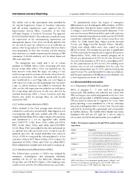
anti-CD34/FITC (1:100, Bioss, USA), rabbit anti-CD44/ Table 1. PCR primer sequences for qRT‑PCR analysis of gene
FITC (1:100, Bioss, USA), and rabbit anti-CD45/FITC expression
(1:100, Bioss, USA) were added to the tube and placed on
ice, and the tube wall was flicked every 10 min to mix the Genes Sequence
antibody and cells. The bound antibodies were added to BMP-2 F: 5’-GATCTGTACCGCAGGCACTC-3’
100 μL of PBS to resuspend the cells and placed in a flow R: 5’-TTCCCACTCATCTCTGGAAGTT-3’
cytometer (Attune NxT, ThermoFisher, USA) to detect the Runx-2 F: 5’-CATTTGCACTGGGTCACACGTA-3’
expression of CD34, CD44, and CD45 on rabbit MSCs. R: 5’-GAATCTGGCCATGTTTGTGCTC-3’
Three independent samples were evaluated in each group. Collagen-1 F: 5’-GGCAATAGCAGGTTCACGTACA-3’
R: 5’-CGATAACAGTCTTGCCCCACTT-3’
2.2.3. Multidirectional differentiation of MSCs SOX9 F: 5’-ACTCCTCCTCCGGCATGAG-3’
R: 5’-GCTGCACGTCGGTTTTGG-3’
The differentiation potential of MSCs was induced by GAPDH-rabbit F: 5’-GGATTTGGCCGCATTGG-3’
osteogenic, chondrogenic, and adipogenic induction R: 5’-CAACATCCACTTTGCCAGAGTTAA-3’
medium. On the 21 day after induction, Alizarin red, BMP-2 and Runx-2 are osteogenesis-specific genes, Collagen-1 and
st
Alcian blue, and Oil Red O (Sigma, USA) stainings were SOX9 are chondrogenesis-specific genes, and GAPDH-rabbit is an
used to detect, whether the differentiation was successful. internal reference gene. PCR: Polymerase chain reaction
Volume 9 Issue 2 (2023) 179 https://doi.org/10.18063/ijb.v9i2.662