
Page 188 - IJB-9-2
P. 188
International Journal of Bioprinting A regulated GelMA-MSCs scaffold by three-dimensional bioprinting
2.3.2. Cell proliferation test incubation at 37°C in 5% CO , cells were harvested for
2
The proliferation ability of MSCs grown on different materials further experiments. Table 2 lists all the microRNA mimics
was detected using the cell counting kit (CCK-8, Beyotime, used in this study, and the experiments were performed in
USA). Different concentrations of GelMA containing MSCs triplicate and repeated in three independent experiments.
were added to the 96-well plates, and CCK-8 detection was 2.4.2. Wound healing assay
carried out on the 1 , 3 , 5 , and 7 days. 10 μL of CCK-8
th
th
st
rd
reagent was added to each well before detection. 100 μL of The wound healing assay was used to detect whether
PBS was loaded to each of the outermost empty wells in the microRNA-410 promotes cell migration. 200 μL sterile
plate to prevent evaporation during the incubation process, pipette tips were used to damage the monolayer cells. After
and the plate was put in the incubator for 2 h. Subsequently, the cell fragments were washed with PBS, the medium
three independent samples were taken and the absorbance was added to the incubator set at 37°C and 5% CO . They
2
at 450 nm was measured with a microplate reader. The were incubated at 0, 6, 12, and 24 h, respectively, and
optimal concentration of GelMA, in which the optimal cells photographed under an inverted microscope. To observe
grow three-dimensionally, was analyzed. the migration of cells at the edge of scratches, ImageJ
software was used to measure the wound area at each time
2.3.3. Live/Dead viability assay point, and three independent experiments were performed.
The viability of cultured cells within the 3D hydrogels was 2.4.3. Transwell migration assay
assessed using the Ca-AM/PI live-dead staining reagent
(Solarbio, China). After equilibrating the Ca-AM and PI Migration ability of MSCs was assessed using the transwell
reagent stock solutions at room temperature for 30 min, migration assay. In this study, we used the Corning Costar
2.5 μL of the PI stock solution was added to 10 mL of PBS, 24-well Transwell Chamber System Assay Kit (8 μm
shaken, and mixed, and then, 5 μL of the Ca-AM stock solution size) to calculate the number of cells that passed through
was added and mixed well. Ca-AM/PI working solution was a polycarbonate membrane. About 200 μL serum-free
5
prepared. Before staining, the adherent cells were gently L-DMEM medium containing 1×10 cells were added to the
washed twice with PBS and discarded to ensure that the active upper chamber. A volume of 500 μL of 10% FBS-containing
esterase contained in the medium was removed. The prepared L-DMEM medium was added to the lower chamber as a
Ca-AM/PI working solution was added to each well of the chemoattractant. After being incubated at 37°C for 48 h, the
24-well plate, and it should be confirmed that the working non-migrating cells on the upper surface were carefully scraped
solution completely covered the scaffold. After incubation off with cotton swabs. Cells that migrated to the bottom of
in a 37°C incubator for 30 min, the staining solution was the membrane were fixed with 4% paraformaldehyde, soaked
aspirated to terminate the incubation. After rinsing twice with PBS, and stained with 1% crystal violet. Stained cells were
with PBS, the plate was put in an incubator set at 37°C and visualized and counted under a microscope. To minimize the
5% CO . The cells were placed on the scaffold for 48 h before bias, three randomly selected fields were quantified, and the
2
staining for living and dead cells. Three independent samples average number of cells was taken. Three experiments were
were photographed for evaluation. Confocal microscope repeated for each group.
(STELLARIS 8, Leica, Germany) images were taken for 2.4.4. Cell proliferation assay
calculating the proportion of living/dead cells.
Ca-AM/PI live-dead staining was used to detect whether
2.4. Regulation of MSCs biological behavior by microRNA-410 promoted cell proliferation. Blank group,
microRNA-410
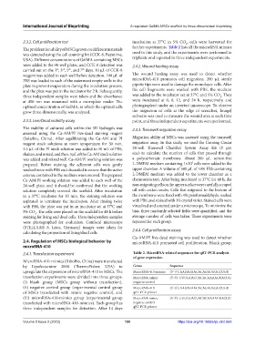
2.4.1. Transfection experiment Table 2. MicroRNA‑related sequences for qRT‑PCR analysis
of gene expression
MicroRNA-410-mimics (RiboBio, China) were transfected
by Lipofectamine 2000 (ThermoFisher, USA) to Genes Sequence
upregulate the expression of microRNA-410 in MSCs. The MicroRNA-410 mimics (5’-3’): AAUAUAACACAGAUGGCCUGU
transfection experiments were divided into three groups: MicroRNA mimic (5'-3'): UUUGUACUACACAAAAGUACUG
(i) blank group (MSCs group without transfection), negative control
(ii) negative control group (experimental control group MicroRNA-410 (5’-3’): AAUAUAACACAGAUGGCCUGU
of MSCs transfected with mimic negative control), and qRT-PCR primer
(iii) microRNA-410-mimics group (experimental group MicroRNA mimic (5'-3'): UUUGUACUACACAAAAGUACUG
transfected with microRNA-410-mimics). Each group has negative control
three independent samples for detection. After 14 days qRT-PCR primer
Volume 9 Issue 2 (2023) 180 https://doi.org/10.18063/ijb.v9i2.662