
Page 192 - IJB-9-2
P. 192
International Journal of Bioprinting A regulated GelMA-MSCs scaffold by three-dimensional bioprinting
migration, proliferation, and differentiation, the growth (**, P < 0.01), most of the cells survived, which fulfills the
of GelMA-60 5% group was better. From the above, requirements of 3D-bioprinted scaffold for transplantation.
preliminary GelMA-60 5% scaffold was selected to repair To observe the 3D growth of GelMA-MSCs bioink
full-thickness cartilage defects in rabbits. more intuitively, the growth of MSCs cultured for 48 h was
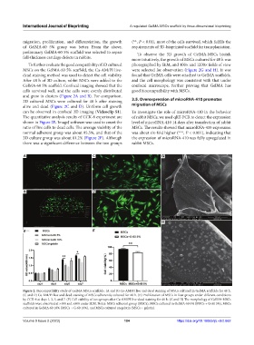
To further evaluate the good compatibility of 3D cultured photographed by SEM, and 400× and 1200× fields of view
MSCs on the GelMA-60 5% scaffold, the Ca-AM/PI live- were selected for observation (Figure 2G and H). It was
dead staining method was used to detect the cell viability. found that GelMA cells were attached to GelMA scaffolds,
After 48 h of 3D culture, rabbit MSCs were added to the and the cell morphology was consistent with that under
GelMA-60 5% scaffold. Confocal imaging showed that the confocal microscope, further proving that GelMA has
cells survived well, and the cells were evenly distributed good biocompatibility with MSCs.
and grew in clusters (Figure 2A and B). For comparison,
2D cultured MSCs were cultured for 48 h after staining 3.5. Overexpression of microRNA-410 promotes
alive and dead (Figure 2C and D). Uniform cell growth migration of MSCs
can be observed in confocal 3D imaging (Videoclip S1). To investigate the role of microRNA-410 in the behavior
The quantitative analysis results of CCK-8 experiment are of rabbit MSCs, we used qRT-PCR to detect the expression
shown in Figure 2E. ImageJ software was used to count the level of microRNA-410 14 days after transfection of rabbit
ratio of live cells to dead cells. The average viability of the MSCs. The results showed that microRNA-410 expression
normal adherent group was about 95.5%, and that of the was about six-fold higher (***, P < 0.001), indicating that
3D culture group was about 81.2% (Figure 2F). Although the expression of microRNA-410 was fully upregulated in
there was a significant difference between the two groups rabbit MSCs.
A C G
B D
H
E F
Figure 2. Biocompatibility study of GelMA-MSCs scaffolds. (A and B) Ca-AM/PI live and dead staining of MSCs cultured in GelMA scaffolds for 48 h.
(C and D) Ca-AM/PI live and dead staining of MSCs adherently cultured for 48 h. (E) Proliferation of MSCs in four groups under different conditions
by CCK-8 at days 1, 3, 5, and 7. (F) Cell viability of two groups after Ca-AM/PI live-dead staining for 48 h. (G and H) The morphology of GelMA-MSCs
scaffolds were observed at ×400 and ×800 under SEM. Notes: MSCs adherent group (MSCs), MSCs cultured in GelMA-60 5% (MSCs + G-60 5%), MSCs
cultured in GelMA-60 10% (MSCs + G-60 10%), and MSCs cultured on gelatin (MSCs + gelatin).
Volume 9 Issue 2 (2023) 184 https://doi.org/10.18063/ijb.v9i2.662