
Page 193 - IJB-9-2
P. 193
International Journal of Bioprinting A regulated GelMA-MSCs scaffold by three-dimensional bioprinting
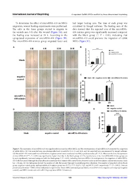
To determine the effect of microRNA-410 on MSCs had larger healing area. The time of each group was
migration, wound healing experiments were performed. calculated by ImageJ software. The healing area of the
The cells in the three groups started to migrate to dots showed that the repaired area of the microRNA-
the scratch area 6 h after the wound (Figure 3A), and 410-mimics group was significantly increased compared
the healing area increased at 24 h. According to the with the blank group (*, P < 0.05), indicating that
upregulated expression of microRNA-410 (Figure 3B). microRNA-410 could promote the migration of rabbit
The microRNA-410-mimics group migrated faster and MSCs (Figure 3C).
A B
C
D E
Figure 3. The expression of microRNA-410 was significantly increased in rabbit MSCs, and the overexpression of microRNA-410 promoted the migration
of rabbit MSCs. (A) The scratched area was photographed and recorded at 0, 6, 12, and 24 h, and the scratched area was measured by ImageJ software.
(B) MicroRNA-410 mimics and microRNA mimic negative control were transfected into MSCs and detected by qRT-PCR. After transfection, the expression
of microRNA-410 (***, P < 0.001) was significantly higher than that in the blank group. (C) The repaired area of the microRNA-410-mimics group can
be seen significantly increased compared with the blank group (*, P < 0.05), indicating that microRNA-410 could promote the migration of rabbit MSCs.
(D) Cells that passed through the polycarbonate membrane were stained with crystal violet, observed under an inverted phase contrast microscope. Scale
bar = 20 μm. (E) The histogram showed the number of cells that passed through Transwell migration chambers. The number of migrated cells in blank
group, negative control group, and microRNA-410-mimics group was 33.75 ± 5.75, 32.25 ± 5.25, and 80.55 ± 6.05, respectively (***, P < 0.001). Data are
presented as the mean ± SEM of three independent experiments. Notes: MSCs untreated group (blank), microRNA mimic negative control group (negative
control), and MSCs transfected with miR-410 group (microRNA-410-mimics).
Volume 9 Issue 2 (2023) 185 https://doi.org/10.18063/ijb.v9i2.662