
Page 197 - IJB-9-2
P. 197
International Journal of Bioprinting A regulated GelMA-MSCs scaffold by three-dimensional bioprinting
A B
Figure 7. Micro-CT scanning and quantitative Analysis. (A) Sagittal cross-sectional 2D images of the femoral condyle of rabbits in each group by micro-CT
scan. (B) Analysis of BV/TV, Tb.Th, Tb.N, and Tb.Sp parameters in subchondral bone defect of femoral condyle of rabbits in each group. Among the three
groups, the GelMA-microRNA-410-MSCs group had the best subchondral bone repair effect, and there was a statistical difference between the 6-week and
12-week groups compared with the former two groups (sample size = 18. *, P < 0.05; **, P < 0.01; ***, P < 0.001).
A existed in the blank group. However, the subchondral bone
defect was filled with a large amount of matrix in the GelMA-
MSCs group, and the position of the new chondrocytes and
collagen was not stretched. In the GelMA-microRNA-410-
MSCs group, the repair effect of the overall subchondral bone
and surface defects was better than that of the blank group
B and the GelMA-MSCs group, and both new collagen and
osteoblasts were densely distributed at the defect repair site,
while the surface defect was not completely repaired. At week
12, there were a few sparse deposits of nascent collagen and
osteoblasts in the blank group. However, the subchondral
C bone defect was filled with more new matrix deposition in
the GelMA-MSCs group. It also can be observed that new
osteoblasts were attached to the periphery of new bone
trabeculae, and new collagen was spread around chondrocytes.
In the GelMA-microRNA-410-MSCs group, the defect repair
effect was the best among the three groups, both surface
cartilage defects and subchondral bone defects were effectively
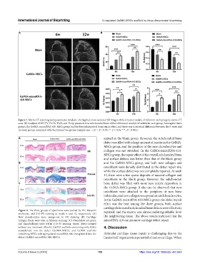
Figure 8. The three groups of specimens were stained by HE, Masson’s repaired, and the matrix was almost indistinguishable from
trichrome, and S-O FS staining at weeks 6 and 12, respectively. (A)
New chondrocytes were orange-red in HE staining. (B) Cartilage the neighboring tissue. The above results indicated that the
collagen fibers were blue in Masson staining. (C) Osteoblasts are green microRNA-410 can promote cartilage defect repair.
and chondroblasts were red in S-O FS staining. Notes: defect sutured
without any treatment (Blank), GelMA scaffolds containing only MSCs 4. Discussion
transplanted into the defect (GelMA-MSCs), and GelMA scaffolds
containing MSCs with upregulated microRNA-410 transplanted into the Articular cartilage tissue repair is challenging due to the
defect (GelMA-microRNA-410-MSCs). limited self-regeneration potential of native cartilage. When
Volume 9 Issue 2 (2023) 189 https://doi.org/10.18063/ijb.v9i2.662