
Page 195 - IJB-9-2
P. 195
International Journal of Bioprinting A regulated GelMA-MSCs scaffold by three-dimensional bioprinting
A B C
D
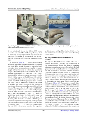
Figure 5. 3D printing process of GelMA-MSCs scaffold. (A and B) The printing process of a 3D extrusion bioprinter. (C and D) The printed cylindrical
scaffold with a height of 5 mm and a diameter of 5 mm.
It was preliminarily shown that GelMA-MSCs bioink proliferation, and cartilage differentiation of MSCs in vitro,
transfected with microRNA-410 had a preliminary effect but also had obvious advantages in the repair of cartilage
on promoting cartilage repair, which may be due to the defects in vivo.
effect of microRNA-410 on cell migration, proliferation,
and differentiation in MSCs, resulting in enhanced repair 3.8. Evaluation and quantitative analysis of
effect. micro-CT
The repair of the rabbit femoral condyle defect can be
As shown in Figure 6C, CT surface reconstruction
technology was used to quantitatively analyze the repaired observed by micro-CT, and the 3D reconstruction of
the NRecon software showed that there are significant
area of the defect surface. Next, the corresponding equal differences among the three groups. The defect in the blank
proportion of the coronal defect was placed in the center. group was barely repaired, and it was revealed that there
ImageJ software was used to perform the quantitative was only minimal to no hard tissue formation within the
analysis. The defect repair ratios at weeks 6 and 12 in defect site throughout the extent of the study. In the GelMA-
the blank group were 9.23 ± 3.41% and 12.34 ± 2.86%, MSCs group, the repaired pore traces could be observed.
respectively. The defect repair ratio at weeks 6 and 12 in the Subchondral bone was irregularly arranged, and the bone
GelMA-MSCs group was 26.45 ± 4.52% and 41.34 ± 4.09%, trabecular density was lower than normal tissue. The
respectively. The defect repair ratio at week 6 and 12 in the density and thickness of the trabecular bone were similar
GelMA-microRNA-410-MSCs group was 54.34 ± 4.32% to the neighboring tissue in the GelMA-microRNA-410-
and 79.65 ± 5.32%, respectively. The statistical process MSCs group (Figure 7A). The quantitative analysis of the
of repairing was carried out in a blinded quantitative statistics was, further, carried out on the basis of NRecon,
analysis, and all data are expressed as mean ± SEM of three CTAn, and Mimics 10.01 software. The subchondral bone
statistics. The repair effect at week 6 in the GelMA-MSCs repair indicators selected in this study are BV/TV, Tb.
scaffold group was better than that in the blank group (*, Th, Tb. N, and Tb. Sp (Figure 7B). In the GelMA-MSCs
P < 0.05), while the GelMA-microRNA-410-MSCs group group and the GelMA-microRNA-410-MSCs group, the
was the best among the three groups (***, P < 0.001). At trabecular bone repair in subchondral bone was better
week 12, the repair effect of the GelMA-MSCs group was than that in the blank group, and their trabecular bone
still significantly better than that of the blank group (**, increased significantly in density, thickness, and volume
P < 0.01), while the GelMA-microRNA-410-MSCs group fraction. In terms of the repair effect of microRNA-410,
had the best repair effect in three groups, reaching a repair the quantitative analysis showed that there were significant
rate of nearly 80%, which was significantly different from statistical differences in the three groups at weeks 6 or 12
the previous group (***, P < 0.001). The results showed (*, P < 0.05; **, P < 0.01; ***, P < 0.001). All of the data were
that microRNA-410 not only promoted the migration, subjected to blinded histological quantitative analysis, and
Volume 9 Issue 2 (2023) 187 https://doi.org/10.18063/ijb.v9i2.662